Structural Molecular Biology
Illuminating Biological Structures at the Atomic and Molecular Levels


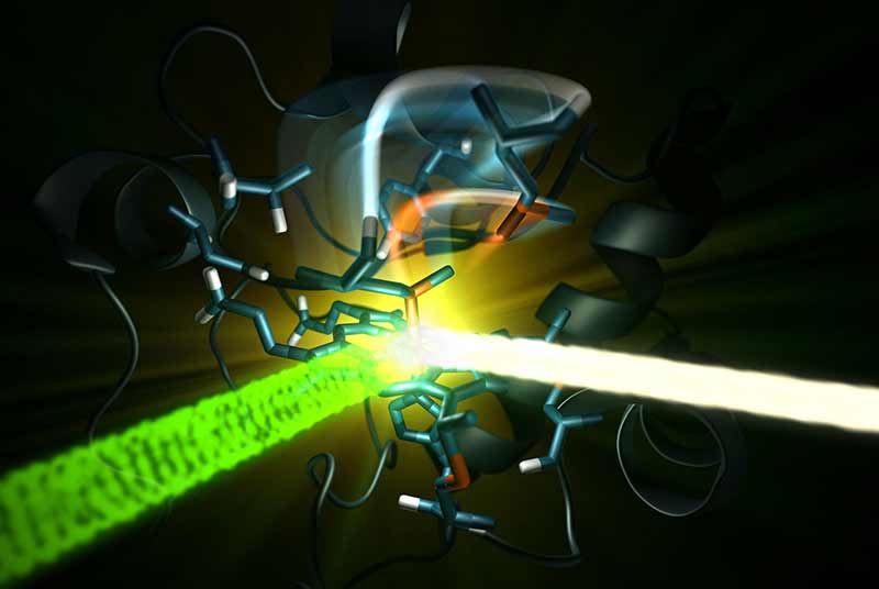
X-ray Spectroscopy & Imaging
SSRL has five hard X-ray Spectroscopy beam lines and three Microfocus Imaging beam lines dedicated or shared for biological, biomedical and bioenergy research funded by the NIH and DOE-BER. The SMB group supports and develops technical instrumentation and theoretical methods for state-of-the-art tender and hard X-ray spectroscopy and EXAFS studies on metalloproteins, cofactors and metals in biology. The SMB group has also contributed to the development of a wiggler end-station beam line and facilities for X-ray emission and Raman spectroscopy, HERFD and RIXS measurements on biological and biomimetic systems. A holistic imaging methodology combining spectroscopy and X-ray fluorescence has been developed on three beam lines encompassing μm-cm scale specimen over the tender & hard X-ray regime integrating hardware and software for seamless measurement across the stations.
SMB Programs
Instrumentation
The SMB group maintains and operates standard and specialized in-hutch instrumentation for a variety of research programs. These include; cryostats for low-temperature measurements, detector systems serving dilute and concentrated samples and specialized instrumentation for tender-energy and single-crystal XAS measurements. Experimental details about beam lines and information about their specific capabilities are listed under each beam lines technical page.
X-ray Absorption Spectroscopy
Advanced Spectroscopy
X-ray Fluorescence Imaging
Software
Data Collection
WebXAS is the SSRL X-ray Absorption Spectroscopy in-browser data acquisition program. It is designed to allow quick and easy XAS experimental setup and data collection, and to make optimal use of available beam time. It has many facilities, including the ability to run remote access, all of which are described in on-line documentation. Click here to see the WebXAS Help Guide. WebGe, included in WebXAS, is used to interface with the multi-element fluorescence detectors at both BL7-3 (30-element Ge detector) and BL9-3 (100-element Ge detector).
Data Analysis
EXAFSPAK and SIXPACK
The data analysis packages EXAFSPAK and SIXPACK are available free of charge. Both are currently installed on the XAS beamline computers. Windows and a variety of Unix versions are currently available. These, and the manual for EXAFSPAK, can be obtained here. The manual for SIXPACK can be obtained here.
The MicroAnalysis Toolkit
The data analysis package, The MicroAnalysis Toolkit, is available free of charge for visualizing and processing X-ray Fluorescence imaging data. The MicroAnalysis Toolkit is available at all of the XRF imaging beamlines, and staff are well versed in assisting users with this software. Instructions for The MicroAnalysis Toolkit download. Basic processing and help for various functions can be found here.
Other Programs
- FEFF: http://leonardo.phys.washington.edu/feff/
- Demeter: https://bruceravel.github.io/demeter/
- IFEFFIT: http://cars9.uchicago.edu/ifeffit/
- GNXAS: http://gnxas.unicam.it/
- SMAK: https://www.sams-xrays.com/smak
Utilities
Some useful utilities to aid user measurements are provided below.
Resources
Groups
- International XAFS Society
- Center For X-Ray Optics At Lawrence Berkeley National Lab
- International Union Of Crystallography
Information/Tutorials
- Grant Bunker's XAFS Tutorial
- Matt Newville's Fundamentals Of XAFS
- EXAFS 2016 - SSRL Summer School on Synchrotron X-ray Spectroscopy
- 2013 SSRL Summer School on Synchrotron X-ray Spectroscopy
Software/Programs
Cross-Section/Absorption Coefficients And Constants
- McMaster Periodic Table Of X-Ray Absorption Values
- Book Of X-Ray Absorption Values
- NIST Attenuation Coeeficient Bibliography
- X-Ray Data Booklet
- XAFS Data Library
- Atoms.Inp Archive
Databases
X-ray Absorption Spectroscopy
X-ray absorption spectroscopy (XAS) is a well-established technique for simultaneous local geometric and electronic structure determination of a metalloprotein active site. XAS is element specific and sample state agnostic, making it ideal for dilute biological solutions. SSRL has two hard x-ray and two tender x-ray biological XAS beamlines, together covering 2-30 KeV. The beamlines are equipped with specialized instrumentation, including low temperature (liq He) cryostats, solid-state Ge and Silicon drift detectors for dilute samples..
Beam Line 9-3
Beam line 9-3 is the side station on the hybrid wiggler beam line 9 dedicated to general user biological XAS. BL9-3 provides high intensity over a broad x-ray energy range, and is designed to provide focused beam with an energy range of 5 keV to 30 keV.
Beam Line 7-3
Beam line 7-3 is the side station on the wiggler beam line 7 dedicated to general user biological XAS. BL7-3 provides medium high intensity over a broad x-ray energy range, and is designed to provide unfocused beam with an energy range of 5 keV to 30 keV.
Beam Line 4-3
Beam line 4-3 is the side station on the wiggler beam line 4 dedicated to biological, environmental and chemical catalysis XAS. BL4-3 provides medium high intensity over a narrow x-ray energy range, and is designed for primarily for studies to the tender x-ray regime.
Beam Line 14-3a
Beam line 14-3a is the side station on the bending magnet beam line 14 dedicated to biological, environmental and chemical catalysis XAS. BL14-3a provides medium high intensity over a narrow x-ray energy range, and is designed for primarily for studies in the tender x-ray regime.
Advanced Spectroscopy
X-ray emission spectroscopy (XES) is a core-level electronic structure determination experimental technique which gives information on orbital splitting, spin and oxidation states as well as the local symmetry and coordination. Beamline 15-2 is a versatile station, equipped with a custom-built, high-resolution analyzer crystal instrumentation, which allows for the measurement of both resonant and non-resonant XES on dilute biological metalloproteins in particular, and metals in biology in general (measured at cryogenic conditions).
Beam Line 15-2
Beam Line 15-2 is an advanced spectroscopy experimental station on the multidisciplinary general user wiggler Beam Line 15 dedicated to X-ray spectroscopy techniques over 5 – 30 KeV. The techniques include X-ray Emission Spectroscopy (XES), Resonant Inelastic X-ray Scattering (RIXS), High Energy Resolution Fluorescence Detection (HERFD) and X-ray Raman Spectroscopy (XRS). Scientific applications are in Structural Biology, Chemical Catalysis and Materials and Environmental Sciences.
X-Ray Emission
X-ray emission spectroscopy (XES) is a core-level electronic structure determination experimental technique which gives information on orbital splitting, spin and oxidation states as well as the local symmetry and coordination. Beam line 15-2 is a versatile station, equipped with a custom-built, high-resolution analyzer crystal instrumentation, which allows for the measurement of both resonant and non-resonant XES on dilute biological metalloproteins in particular, and metals in biology in general (measured at cryogenic conditions).
RIXS
Resonant inelastic x-ray scattering (RIXS) is a spectroscopic technique, which uses monochromatic incident and emitted fluorescent x-rays to create a 2p-3d core emission spectral intensity map, which holds electronic information simultaneously about the bound and valence energy levels of the absorbing atom, although the information is distinct from that obtained from the individual absorption and emission processes involved in RIXS. Beam line 15-2 is equipped with a cryostat to perform RIXS on sensitive biological systems.
HERFD
The X-ray absorption near-edge region holds subtle electronic structure information about orbital splitting, spin and oxidation states. However, standard x-ray absorption spectra are broadened due to the large finite lifetime of the core hole, much broader than that of the valence levels being probed. Using only a single fluorescence line with energy resolution better than the core hole life time results in the measurement of high-resolution fluorescence detected XANES spectra. This technique is especially important for high Z element with large core hole life times. Beam line 15-2b is equipped with analyzer crystals that allow for HERFD measurements on biological systems.
X-Ray Raman
(XRS) is the non-resonant inelastic scattering of x-rays from core electrons. It is analogous to Raman scattering, which is a widely used tool in optical spectroscopy, with the difference being that the wavelengths of the exciting photons fall in the x-ray regime and the corresponding excitations are from deep core electrons. XRS is an element-specific spectroscopic tool for studying the electronic structure of matter. In particular, it probes the excited-state density of states (DOS) of an atomic species in a sample. On BL 15-2 XRS is used as a powerful hard x-ray method to probe the soft x-ray region without the constraints of vacuum environment.
X-ray Fluorescence Imaging
X-ray fluorescence imaging utilizes the high brightness of SPEAR3 and focused beam generated by the uses of Sigray achromatic paraboidal lenses, capillaries and apertures to study spatial distribution of elements in biological samples such as brain tissue. The imaging beam lines have the unique capability of combining spatial mapping with chemical and structural information of various elements through XAS (edges and EXAFS). The three beam lines are equipped with standardized instrumentation for seamless measurement across 2-30 eV energy range and 1-250 µm length scales, as well as cryostat stage capabilities for imaging cold tissues available at BL2-3 and BL14--3. Additional sample preparation laboratory facilities include microtome/cryotome, optical microscopy and FTIR.
Beam Line 2-3
BL2-3 is a dedicated micro-XAS bending magnet beam line with sigray achromatic paraboidal lensesas the focusing optics to achieve a spot size of 1 or 5 µm2. The only optical element in front of the experimental hutch is a water-cooled variable-exit monochromator. The presence of minimal optical elements upstream of the experimental focusing optics imparts exceptional spatial stability to the micro-focused spot. Experiments conducted on this beam line include XRF imaging, micro-XAS (both XANES and EXAFS), XANES imaging, micro-diffraction and fluorescence tomography.
Beam Line 7-2
BL7-2 (formerly BL10-2a) is high flux wiggler beamline dedicated to large format (600 x 300 mm) XAS imaging. A combination of capillary optics and pinhole apertures are used to achieve beam sizes from ~25 to 250 µm2. The higher flux on beamline 10-2b (~10-50 times greater than BL 2-3) provides a complementary set of abilities to BL 2-3, such as rapid scanning over larger areas and the ability to detect lower elemental concentrations. Experiments conducted on this beam line include XRF imaging, micro-XAS (both XANES and EXAFS) and XANES imaging. Change the link to BL7-2 when for view details button
Beam Line 14-3b
Beam line 14-3b is a bending magnet side station dedicated to X-ray imaging and micro X-ray absorption spectroscopy of biological, biomedical, materials, and geological samples. 14-3b is equipped with specialized instrumentation for XRF imaging data collection in the tender X-ray regime of 2-5 KeV. Sigray achromatic paraboidal lenses are used to achieve microfocus beam of 1 or 5 µm2. BL14-3b is ideal for obtaining elemental maps of small samples (~ 20 µm to 25 mm) as well as performing XANES on selected points in the sample (1 or 5 µm2). Experiments conducted on this beam line include XRF imaging, micro-XAS and XANES imaging.