You are here

BL4-2

Quantifying Myelin and Axon Orientations in the Brain

Myelin is an indispensable component of our brain, “insulating” neurons and enabling fast signal transduction. The level of myelination and the organization of myelinated axons are important determinants of brain development and are affected in virtually all neurological diseases. However, current myelin imaging methods in clinics or research are either non-specific (e.g. MRI) or destructive (e.g. histology). Small-angle x-ray scattering (SAXS), in contradistinction, combines the high tissue penetration of x-rays with specificity to periodic tissue nanostructures.

Figure 1. a) In SAXS-TT, the x-ray beam raster scans the sample under different rotations and tilts. b) One projection of the mouse brain, with the myelin peak signal intensity, and 2D fiber orientation color-encoded. c-d) Tensor tomographic reconstruction of the myelin-specific signal results in a tensor representing the myelinated axon orientation distribution per voxel (d) and quantitative 3D myelin maps (c).

In this study, the recently developed method of small-angle x-ray scattering tensor tomography1 (SAXS-TT) was used for deriving myelin levels and axon orientations in the central and peripheral nervous system. By exploiting the nano-structural ~17 nm repeat pattern typical of myelin sheath, which gives rise to a distinct small angle scattering pattern, tomographic myelin maps in macroscopic tissue volumes were derived (Fig. 1). The method was demonstrated on whole mouse brain, mouse spinal cord and human white matter samples.

The ability of the method to provide accurate myelin level readouts was assessed using myelin histology as gold standard. In all cases the SAXS-TT derived myelin levels were very highly correlated with histology results; moreover, when compared to myelin-sensitive MRI contrasts, histology and SAXS-TT gave the same output, suggesting SAXS-TT’s potential as reference myelin imaging method, with the additional benefits of no extra sample preparation, its nano-structural specificity and its non-destructive nature.

Similar results were obtained for the axonal orientation maps, which correlated highly with both MRI measures and 3D histology. Since MRI metrics are not specific to myelinated axons, and histology is destructive and non-quantitative, this study suggests that SAXS-TT has the potential to become the reference method for assessing myelinated axon orientations, responsible for long-range connectivity.

The excellent sensitivity and accuracy of the method was further showcased in a variety of mouse and human samples of different sizes. In a sample of mouse spinal cord, it was shown that neural tracts derived with SAXS-TT reflect the neural tracts more accurately than the ones derived from diffusion MRI, which is the current method of choice for the 3-dimensional mapping of neural tracts.

The sensitivity of the SAXS-TT method to small differences in periodicity of the myelin structure also enables it to distinguish between the central and peripheral nervous system (CNS/PNS) myelin. This allows for multiplexed imaging, where CNS and PNS myelin signals could be analyzed separately enabling the first 3-dimensional rendering of their transition areas between the central and peripheral nervous system, revealing a ball-socket fit.

It was further demonstrated that the method can be applied to study neurologic diseases by examining normal and dysmyelinated mouse brains, showing very different myelin levels as well as altered myelin periodicity in the diseased brain, emphasizing the potential of the SAXS-TT based myelin mapping as a future diagnostic tool.

SAXS-TT experiments for this study were performed at the BioSAXS BL4-2 at the Stanford Synchrotron Radiation Lightsource (SSRL), the cSAXS beamline of the Swiss Light Source (SLS) as well as the LiX beamline at the National Synchrotron Light Source II (NSLS-II).

In summary, this novel non-destructive, stain-free imaging approach enables quantitative studies of myelination within and across nervous tissue samples during development, aging, disease and treatment. It provides highly accurate maps of myelin distribution and axon orientation in macroscopically sized samples. It should be noted that the general method is also applicable to other ordered biomolecular nanostructures and such extensions are currently being investigated.

Primary Citation(s): 
M. Georgiadis, A. Schroeter, Z. Gao, M. Guizar-Sicairos, M. Liebi, C. Leuze, J. A. McNab, A. Balolia, J. Veraart, B. Ades-Aron, S. Kim, T. Shepherd, C. H. Lee, P. Walczak, S. Chodankar, P. DiGiacomo, G. David, M. Augath, V. Zerbi, S. Sommer, I. Rajkovic, T. Weiss, O. Bunk, L. Yang, J. Zhang, D. S. Novikov, M. Zeineh, E. Fieremans and M. Rudin, "Nanostructure-specific X-ray Tomography Reveals Myelin Levels, Integrity and Axon Orientations in Mouse and Human Nervous Tissue", Nat. Commun. 12, 2941 (2021) doi: 10.1038/s41467-021-22719-7
References: 

M. Liebi, and M. Georgiadis et al., “Nanostructure Surveys of Macroscopic Specimens by Small-angle Scattering Tensor Tomography”, Nature 527, 349 (2015).

Beamlines Used:

Topic:

Cross-β Structure - a Core Building Block for Streptococcus mutans Functional Amyloids

Most amyloids1 are misfolded proteins, having enormous variety in native structures. Pathological amyloids are implicated in diseases including Alzheimer’s disease and many others.  They are characterized by long, unbranched fibrillar structure, enhanced birefringence on binding Congo red dye, and cross-β structure – β-strands running approximately perpendicular to the fibril axis, forming long β-sheets running in the direction of the axis.  Fiber diffraction patterns from amyloids are marked by strong intensity at about 4.8 Å in the meridional direction (parallel to the fibril axis), corresponding to the separation of strands in a β-sheet, and in many cases broader but distinct equatorial intensity at about 10 Å.  The 10 Å intensity (whose position may vary considerably) comes from the distance between stacked β-sheets.  This stacking is characteristic of the many amyloids formed by small peptides, including peptide fragments of larger amyloidogenic proteins.  While some authors have required the 10 Å intensity to characterize an amyloid, it is not strictly necessary, since architecturally more complex examples have been found of Congo-red-staining fibrils with cross-β structure, but without the stacked-sheet structure, and consequently without the 10 Å intensity on the equator.


Figure 1. Crystal or predicted 3D structures of S. mutans
C123 (left), AgA (center), and Smu_63c (right).

Amyloids do not always stem from protein misfolding.  Organisms across all kingdoms utilize functional amyloids in numerous biological processes.  Bacteria are no exception. Bacterial amyloids contribute to biofilm formation and stability.  Tooth decay is the most common infectious disease in the world.  A major etiologic agent, Streptococcus mutans, is a quintessential biofilm dweller that produces at least three different amyloid-forming proteins, adhesins P1 and WapP, and the cell density and competence regulator Smu_63c2.  The naturally occurring truncation derivatives of P1 and WapA, C123 and AgA, represent the amyloidogenic moieties, and a new paradigm of Gram-positive bacterial adhesins is emerging of adhesins having dual functions in monomeric and amyloid forms. While each S. mutans protein possesses considerable β-sheet structure, the tertiary structures of each protein are quite different (Fig. 1).  This study further characterized S. mutans amyloids and addressed the ongoing debate regarding the underlying structure and assembly of bacterial amyloids including speculation that they are structurally dissimilar from better-characterized amyloids.


Figure 2. Transmission Electron Microscopy (left) and x-ray fiber diffraction patterns (right) of S. mutans AgA amyloid in mat (top) and fiber form (bottom).


The highlighted work utilized classical x-ray fiber diffraction performed at the SSRL biological SAXS Beam Line 4-2 to characterize S. mutans amyloids that display an incompletely understood mat-like structure. During the course of sample preparation and protease digestion to remove residual monomers from amyloid preparations of purified proteins, it became apparent that typical amyloid fibers were visualized after proteolysis, and that mats were reconstituted by incubation of monomers with fibers.  When microbiologists at the University of Florida were introduced by SSRL Beam Line 4-2 staff scientists to structural biologists from Vanderbilt University, the question of S. mutans basic amyloid structure was answered.  In a series of synchrotron based fiber diffraction experiments at SSRL Beam Line 4-2, the research team established for the first time that the classical β-amyloid structure was indeed the core building block of both amyloid mats and fibers for all three bacterial amyloid-forming proteins.  AgA is shown here as an example (Fig. 2).

The establishment of a conserved fiber diffraction pattern of S. mutans amyloids will enable future studies to evaluate biofilm matrix material of this and other microbes.  This method and structural foundation can now be employed to assess the impact of varying environmental parameters on amyloid assembly within biofilms, and of potential treatment of biofilm-dependent diseases by amyloid inhibitors.  Sometimes old-school is the most valuable tool!

Primary Citation(s): 
A. L. Barran-Berdon, S. Ocampo, M. Haider, J. Morales-Aparicio, G. Ottenberg, A. Kendall, E. Yarmola, S. Mishra, J. R. Long, S. J. Hagen, G. Stubbs and L. J. Brady, "Enhanced Purification Coupled with Biophysical Analyses Shows Cross-β Structure as a Core Building Block for Streptococcus mutans Functional Amyloids", Sci. Rep. 10, 5138 (2020)
References: 
  1. G. Stubbs and J. Stöhr, “Structural Biology of PrP Prions”, Cold Spring Harb. Perspect. Med. 7, 113 (2017)
  2. R. N. Besingi, I. B. Wenderska, D. B. Senadheera, D. G. Cvitkovitch, J. R. Long, Z. T. Wen, L. J. Brady, “Functional Amyloids in Streptococcus mutans, Their Use as Targets of Biofilm Inhibition and Initial Characterization of SMU_63c.”, Microbiology 163, 488 (2017)

Beamlines Used:

Topic:

Structure of the Full-length Clostridium difficile Toxin B

August 2019

Clostridium difficile (C. diff) is an opportunistic pathogen that establishes in the colon when the gut microbiota are disrupted, often seen in seriously ill or elderly patients.  Clostridium difficile infection (CDI) has become the most common cause of antibiotic-associated diarrhea and gastroenteritis-associated death in developed countries, accounting for a half-million cases and 29,000 deaths annually in the US. It is considered an “urgent threat” by the CDC.

The pathology of CDI is primarily mediated by homologous exotoxins, TcdA and TcdB. While the relative roles of these two toxins in the pathogenesis of CDI are not completely understood, recent studies have shown that TcdB is more virulent than TcdA and more important for inducing the host inflammatory and innate immune responses.
 

Figure. 1. Overall structure of the full length TcdB holotoxin. (a) A schematic diagram showing the domain organization of TcdB and the approximate VHH-binding regions. GTD, glucosyltransferase domain (red); CPD, cysteine protease domain (light blue); DRBD, delivery and receptor-binding domain (yellow); CROPs, combined repetitive oligopeptides domain (blue). (b) Cartoon representations of TcdB holotoxin. The 3 VHHs that were used to facilitate crystallization were omitted for clarity. The TcdB domains are colored the same as that shown in panel (a).
 

Both TcdB and TcdA are large molecular weight proteins with multiple domains and complicated conformational flexibility. Numerous structures have been reported for fragments of TcdA and TcdB, which have provided tremendous insight into the functions of these toxin domains. However, it remains unknown how individual domains interact within the supertertiary structure of the holotoxin and how the holotoxin dynamically responds in a precise stepwise manner to the environmental and cellular cues that lead to intoxication.

A recent study, led by researchers from the University of California, Irvine (UCI), uncovered the long-sought-after, three-dimensional structure of a toxin primarily responsible for devastating Clostridium difficile infection (Fig. 1). Published in Nature Structural & Molecular Biology, the study sheds light on the weaknesses of TcdB, one of the toxins secreted by the Clostridium difficile bacteria and the main cause of CDI.

The team determined the crystal structure of the toxin by collecting macromolecular crystallography data at the Northeastern Collaborative Access Team 24-ID-C beam line at the Advanced Photon Source (APS) and at Beam Line 9-2 at the Stanford Synchrotron Radiation Lightsource (SSRL). To further probe the structural dynamics of the toxin, the team used a combination of small-angle x-ray scattering (SAXS), single-molecule fluorescence resonance energy transfer (smFRET), and cross-linking mass spectrometry (XL-MS), of which the small-angle x-ray scattering experiments were performed at SSRL Beam Line 4-2.

This study revealed the 3D structure of the gigantic TcdB holotoxin at a near atomic resolution for the first time and indicated the pH-dependent structural flexibility of TcdB that may help to modulate its activity in response to environmental pH change.  through this studty the team also demonstrated how three antibodies could neutralize TcdB, revealing intrinsic vulnerabilities of the TcdB toxin that could be exploited to develop new therapeutics and vaccines for the treatment of CDI.

Standard treatment for CDI involves using broad spectrum antibiotics that reduce the level of C. diff bacteria.  However, the treatment also kills the good bacteria in the gut and disrupts the normal gut microbiome. This approach often leads to frequent disease recurrence.

More potent and cost-effective therapies for CDI remain a desperate need. The 3D structure of TcdB literally provides a blueprint for the development of next-generation vaccines and therapeutics that have enhanced potency and broad-reactivity across different C. diff strains. The UCI team has already begun working on a novel vaccine based on the new structure with early studies showing promising results.
 

Primary Citation(s): 
P. Chen, K.-h. Lam, Z. Liu, F. A. Mindlin, B. Chen, C. B. Gutierrez, L. Huang, Y. Zhang, T. Hamza, H. Feng, T. Matsui, M. E. Bowen, K. Perry and R. Jin, "Structure of the Full-length Clostridium difficile Toxin B", Nat. Struct. Mol. Biol. 26, 712 (2019) doi: 10.1038/s41594-019-0268-0 (link is external)

Beamlines Used:

Topic:

Releasing the Brakes on Apoptosis: Peptide Antagonists Trigger Dimerization and Autoubiquitination of Cellular Inhibitor of Apoptosis Protein 1

January 2012

Programmed cell death, or apoptosis, is a critical failsafe against uncontrolled proliferation.  For this reason, apoptosis is frequently defective in cancer cells, allowing tumor growth to proceed unchecked.  The inhibitor of apoptosis proteins, or IAPs, are some of the final “brakes” on apoptosis, directly inhibiting both caspases and their upstream activators (1,2,3,4).  Thus it is unsurprising that IAP proteins are over-expressed in many human cancers (2,5).

Crystal structure of the monomeric form of cIAP1-B3R.
Figure 1 A) Domain structure of the cIAP1-B3R construct. B) Crystal structure of the monomeric form of cIAP1-B3R. Individual domains are colored as in the primary structure diagram at top. Linkers between domains are colored yellow. The RING domain (orange), which is responsible for homodimerization, is sequestered on three sides by the other globular domains.

The anti-apoptotic functions of IAP proteins are mitigated by the second mitochondrial activator of caspases (SMAC) (6,7).  The N-terminal tail of SMAC, comprising the amino acid sequence AVPI, interacts with a peptide binding pocket on IAP proteins, preventing their association with caspases (8,9,2,4) and triggering the degradation of cellular IAP1 (cIAP1) (10-12).

cIAP1 (like several other IAP family members) is a ubiquitin ligase.  It comprises three baculovirus IAP repeat (BIR) domains, a ubiquitin association (UBA) domain, a caspase association and recruitment domain (CARD), and a RING E3 ligase domain.  A shortened construct consisting of only the BIR3-RING domains (cIAP1-B3R) is sufficient for antagonist-induced activation of ligase activity.  Upon binding of the SMAC peptide or peptide mimetics to the BIR3 domain, we observed that cIAP1-B3R forms a RING-based dimer in vitro.  This dimer form of the protein is active for ubiquitin ligation and leads to cIAP1’s auto-ubiquitination and subsequent degradation by the proteasome.  In this way, the release of SMAC from mitochondria abrogates the anti-apoptotic activity of cIAP1.

To understand the mechanism by which binding of a short peptide at the BIR3 domain triggers dimerization at the 300-amino-acid distant RING domain, we solved the structures of the monomeric and dimeric forms of cIAP1-B3R by X-ray crystallography and small angle X-ray scattering, respectively.  Unexpectedly, the monomeric form of cIAP1 (Figure 1) adopts a wrapped conformation in which the RING domain is sequestered by the BIR3, UBA and CARD domains.  This arrangement prevents the RING domain from adopting the conformation needed for homodimerization.  Targeted mutagenesis of residues at the interface of the RING and BIR3 domains resulted in dimerization of cIAP1 even in the absence of peptide antagonists, confirming the inhibitory role of this conformation.

Ab initio SAXS envelopes of the dimeric forms of MBP-tagged cIAP1-B3R, cIAP1-B3R, and cIAP1 UBA-RING.
Figure 2 Ab initio SAXS envelopes of the dimeric forms of MBP-tagged cIAP1-B3R, cIAP1-B3R, and cIAP1 UBA-RING. Note the planar, extended shape of each construct. Proposed models for each construct are shown at right. Asterisks indicate bound antagonist.

We were able to solve the structure of three dimeric fragments of cIAP1 using small angle X-ray scattering (SAXS): a UBA-RING truncation, cIAP1-B3R, and cIAP1-B3R with an N-terminal maltose binding protein (MBP) tag.  Ab initio model calculation on all three of these constructs revealed extended, nearly planar dimeric structures (Figure 2).  The long, flexible linkers connecting the globular domains of cIAP1 made the creation of a molecular model from these data challenging.  Nevertheless, the relative size and shape of these structures made it clear that the RING-RING dimer is at the center of the complex, with the CARD, UBA and BIR3 domains extended out in a splayed “V.”

The dramatically different monomeric and dimeric structures of cIAP1-B3R, along with in vitro and cellular data demonstrating the functional activity of the dimer, have allowed us to construct the following model for antagonist-based degradation of cIAP1.  Initially, the protein exists in a compact, wrapped form incapable of dimerization.  Upon disruption of the BIR3:RING interface—by SMAC peptide binding, drug binding, or mutation—the monomer unwraps, allowing RING:RING dimerization to occur.  This form of the protein is active for ubiquitin ligation, and it rapidly ubiquitinates itself and is degraded by the proteasome.  Apoptosis follows.  Our understanding of cIAP1’s autoubiquitination mechanism has significant implications for the development of anti-cancer therapeutics aimed at releasing the brakes on apoptosis.

A model for antagonist-induced dimerization, activation, and degradation of cIAP1
Figure 3 A model for antagonist-induced dimerization, activation, and degradation of cIAP1. In monomeric form, cIAP1-B3R exists in the closed, compact conformation. Binding of antagonist to the BIR3 domain disrupts the BIR3:RING interface, causing the compact structure to unwrap, making the RING domain available for dimerization. The dimeric form of the protein is an active ubiquitin ligase. It ubiquitinates itself, leading to its degradation by the proteasome.

Crystallography data was collected at the Advanced Light Source (ALS) and the Advanced Photon Source(APS).  SAXS data was collected at the Stanford Synchrotron Radiation Lightsource (SSRL) and the Advanced Light Source. The ALS is supported by the Director, Office of Science, Office of Basic Energy Sciences, of the U.S. Department of Energy (DOE) under contract no. DE-AC02-05CH11231. SSRL is a Directorate of SLAC National Accelerator Laboratory and an Office of Science User Facility operated for the DOE Office of Science by Stanford University. The SSRL Structural Molecular Biology Program is supported by the DOE Office of Biological and Environmental Research and by the Biomedical Technology Program, National Center for Research Resources, NIH (P41RR001209). Use of the APS, a facility operated for DOE Office of Science by Argonne National Laboratory, was supported under contract no. DE-AC02-06CH11357.

Primary Citation(s): 
E. C. Dueber, A. J. Schoeffler, A. Lingel, J. M. Elliott, A. V. Fedorova, A. M. Giannetti, K. Zobel, B. Maurer, E. Varfolomeev, P. Wu, H. J. Wallweber, S. G. Hymowitz, K. Deshayes, D. Vucic, W. J. Fairbrother. Antagonists Induce a Conformational Change in cIAP1 That Promotes Autoubiquitination. Science 334, 376 (2011).
References: 

1. G. S. Salvesen, C. S. Duckett, Nat. Rev. Mol. Cell Biol.3, 401 (2002).

2. D. Vucic, W. J. Fairbrother, Clin. Cancer Res.13, 5995 (2007).

3. G. S. Salvesen, J. M. Abrams, Oncogene23, 2774 (2004).

4. J. N. Dynek, D. Vucic, Cancer Lett.(2010).

5. A. M. Hunter, E. C. LaCasse, R. G. Korneluk, Apoptosis12, 1543 (2007).

6. C. Du, M. Fang, Y. Li, L. Li, X. Wang, Cell102, 33 (2000).

7. A. M. Verhagen et al., Cell102, 43 (2000).

8. Z. Liu et al., Nature408, 1004 (2000).

9. G. Wu et al., Nature408, 1008 (2000).

10. A. Gaither et al., Cancer Res.67, 11493 (2007).

11. E. Varfolomeev et al., Cell131, 669 (2007).

12. J. E. Vince et al., Cell131, 682 (2007). 

Beamlines Used:

Topic:

Structural Basis for Iron Piracy by Pathogenic Neisseria

January 2013

Of the 11 species of Neisseria bacteria that colonize humans, 9 of them coexist peacefully with us. However,two can cause serious disease N. gonorrhoeae, responsible for the sexually transmitted disease gonorrhea, and N. meningitidis, which causes septicemia and meningitis.  While both pathogenic species can lead to significant long-term consequences, normally only N. meningitidis causes significant fatalities. Onset of infection is quick, resulting in about a 15% mortality rate and leaving 20% of those who survive with lifelong complications.  Antibiotics are prescribed against these pathogens post-infection, but increasing resistance has spurred an immediate push for vaccine development (1).

Commercially available vaccines exist that work against four of the five disease-causing serogroups of N. meningitidis that have been isolated (A, B, C, Y, W135) but there is no vaccine against serogroup B (menB), nor is there a vaccine available against N. gonorrhoeae. One approach for vaccine development against menB and N. gonorrhoeae is to target the iron transporters found on the surface of the pathogens*.  Without iron, these pathogens cannot survive; therefore the transporters are usually well-conserved across Neisserial strains and generally do not undergo significant genetic variation, making them ideal targets for vaccine development.

Pirating Neisseria fig1
Figure 1. Interactions between Neisserial TbpA and human transferrin. a. The crystal structure of Neisserial TbpA (β-barrel domain in green, plug domain in red) in complex with human transferrin (N-lobe shown in pink and C-lobe in gold). Only the C-lobe interacts with TbpA. b. Zoomed view of the interactions between the TbpA helix finger and plug loop with transferrin. c. Molecular interactions between the TbpA helix finger with transferrin. d. Alignment of conformations of the C-lobe of transferrin comparing apo, holo, and TbpA-bound states. TbpA locks the C-lobe of transferrin in a conformation roughly halfway between that observed for apo and holo. e. Proposed mechanism for how TbpA catalyzes the release of iron from transferrin. TbpA residue K359 is believed to hijack the pH sensing triad mechanism of transferrin which leads to repulsion between transferrin residues K534 and R632 and a conformational change that releases iron.

In this study, the crystal structures of two of the surface receptors of menB, TbpA and TbpB, were determined.  These receptors are used specifically by Neisseria to pirate iron from the abundant human iron binding protein, transferrin, during pathogenesis.  TbpA is an essential TonB-dependent transporter (22-stranded b-barrel membrane protein) that is responsible for transporting iron across the outer membrane.  Remarkably, this structure was crystallized in complex with human transferrin, which allowed a precise description of the interactions between the Neisserial TbpA and the human transferrin protein (Fig. 1a), enabling the identification of a helix finger and plug loop that are crucial for function (Fig. 1b,c).  Neisserial TbpA was seen to lock transferrin in a slightly open conformation, sufficient to allow iron release from the cleft (Fig. 1d).  These results led to a plausible mechanism for how Neisserial TbpA can catalyze the release of iron from transferrin at neutral pH for internalization (Fig. 1e).  Further, antibodies based on this structure were developed against Neisserial TbpA that could effectively block transferrin binding using in vitro assays.  MD simulations designed to mimic interactions with the Ton system also revealed a mechanism for how the iron, once extracted, is transported across the outer membrane through the b-barrel domain of TbpA.

Also reported in this study was the structure of Neisserial TbpB (Fig. 2a), a lipoprotein co-receptor that significantly increases the efficiency of iron uptake by specifically binding only iron-containing transferrin and concentrating it on the Neisserial surface.  TbpB then shuttles the iron-loaded transferrin to TbpA which subsequently extracts and imports the iron across the Neisserial outer membrane.  While attempts to crystallize the TbpB-transferrin complex were unsuccessful, SAXS analysis based on data collected at the BL4-2 beam line at SSRL was instrumental in constructing a model for how the Neisserial co-receptor was able to interact with human transferrin at the cell surface (Fig. 2b), revealing that TbpA and TbpB could simultaneously bind transferrin at distinct sites (Fig. 2c).  The SAXS model was further verified when Calmettes, et al. later reported the structure of the TbpB-transferrin complex using X-ray crystallography.

Pirating Neisseria fig 2
Figure 2. Models for the Neisserial TbpB and human transferrin complex and for the fully assembled iron import complex from Neisseria. a. The crystal structure of Neisserial TbpB (purple) aligned to known TbpB structures from porcine pathogens. Residues shown to affect transferrin binding are indicated and were used when modeling the complex with transferrin. b. SAXS analysis was used to model the TbpA-transferrin complex. Shown here is the model fit into the SAXS envelope. c. TbpA (green) and TbpB (cyan) were seen to have distinct non-overlapping binding sites on transferrin. d. Based on the x-ray crystallography and SAXS model, a model for the fully assembled iron import complex was formed (TbpA in green, TbpB in cyan, and transferrin in gold). e. EM analysis of the fully assembled iron import complex was shown to be consistent with our model (panel d).

Based on the X-ray crystallography and SAXS results, the fully assembled Neisserial transferrin-iron import complex (TbpA-TbpB-transferrin) was modeled (Fig. 2d) and EM studies performed to look at it experimentally.  Here, purified complex was used for negative-stain EM analysis to produce class averages of the complex (Fig. 2e).  These class averages were found to be consistent with the model for the fully assembled Neisserial transferrin-iron import complex, allowing the first look at what the transferrin-iron import complex looks like at the Neisserial surface during its pathogenesis.

Primary Citation(s): 
N. Noinaj, N. C. Easley, M. Oke, N. Mizuno, J. Gumbart, E. Boura, A. N. Steere, O. Zak, P. Aisen, E. Tajkhorshid, R. W. Evans, A. R. Gorringe, A. B. Mason, A. C. Steven, S. K. Buchanan, “Structural Basis for Iron Piracy by Pathogenic Neisseria”. Nature 483, 53 (2012). doi: 10.1038/nature10823 (link is external)
References: 

C. Calmettes, J. Alcantara, R. H. Yu, A. B. Schryvers, T. F. Moraes, "The structural basis of transferrin sequestration by transferrin-binding protein B", Nat. Struct. Mol. Biol. 19, 358 (2012)

Beamlines Used:

Topic:

The Structure and Dynamics of Eukaryotic Glutaminyl-tRNA Synthetase

May 2013

Aminoacyl-tRNA synthetases are required in all three domains of life to add the correct amino acid to its cognate tRNA, an essential step in the process of protein synthesis. In eukaryotes and some bacteria, the traditional pathway of aminoacylation exists for glutamine, in which glutaminyl-tRNA synthetase (GlnRS) binds to tRNAgln, glutamine and ATP and first forms a glutaminyl adenylate molecule that is then covalently attached to the 3’-end of tRNAgln with the release of AMP. In most bacteria and all archaea, a different pathway exists where a non-discriminating glutamyl-tRNA synthetase (GluRS) attaches glutamic acid to both tRNAglu and tRNAgln. The misacylated glu-tRNAgln is then converted to gln-tRNAgln by the GatCAB amidotransferase enzyme in bacteria and some archaea, or by the GatDE amidotransferases in other archaea.


Full-length Gln4 shown bound to tRNAgln.

The eukaryote Saccharomyces cerevisiae contains a specific glutaminyl-tRNA synthetase (ScGlnRS), which is an 809-residue protein that contains the signature motifs found in all Class I tRNA synthetases as well as a 215-residue domain appended to its N-terminus. This domain is nearly ubiquitous among eukaryotic GlnRS species, but is absent in prokaryotic homologs; indeed, eukaryotic tRNA synthetases have often been shown to contain additional domains appended to their N-terminal or C-terminal ends, compared to their prokaryotic homologs.  Some of these domains are known to be involved in various roles, including nucleic acid binding, protein-protein interactions, and hydrolytic editing mechanisms, but the functions of many remain uncertain.

Using remotely collected data from SSRL a research group led by Edward Snell of the Hauptman-Woodward Medical Research Institute previously described the structure of the N-terminal domain (NTD) of ScGlnRS, revealing that it has an extraordinary structural resemblance to the region of the B subunit of the GatCAB amidotransferase that binds to tRNAgln. Although structural data for two prokaryotic GlnRS species exists, no structure has been reported for any full-length eukaryotic GlnRS. Using SSRL macromolecular crystallography Beam Line 11-1 Snell's group also determined the structure of the C-terminal domain (CTD) of ScGlnRS from crystals of full-length GlnRS. Based on this structure, on the structure of the NTD, and on small angle x-ray scattering (SAXS) data of the full-length enzyme measured at SSRL Beam Line 4-2, they have developed a model of the full-length enzyme in solution.  Using crystallographic structures and homology with known transamidosome and GlnRS-tRNA complex structures, they also modeled the full-length enzyme bound to tRNAgln . The combined results suggest that C-terminal domain binding to tRNA results in a large conformational reorientation of the N-terminal domain allowing for interactions between the N-terminal domain and the tRNA. The N-terminal domain plays a direct role in tRNA binding.

The combination of crystallographic and solution SAXS studies enabled by SSRL facilities has yielded fundamental new insights into the structural rearrangements occurring in eukaryotic GlnRS-tRNAgln complex formation.

Primary Citation(s): 
T. D. Grant, J. R. Luft, J. R. Wolfley, M. E. Snell, H. Tsuruta, S. Corretore, E. Quartley, E. M. Phizicky, E. J. Grayhack and E. H. Snell, "The Structure of Yeast Glutaminyl-tRNA Synthetase and Modeling of Its Interaction with tRNA", J. Mol. Biol. (2013) doi: 10.1016/j.jmb.2013.03.043

Beamlines Used:

Topic:

Architectures of Whole-module and Bimodular Proteins from 6-Deoxyerythronolide B Synthase

July 2014

Secondary metabolites produced by microorganisms have a market value of over $30 billion annually, and nearly half of these compounds are naturally produced by bacteria in the phylum Actinobacteria1. Phylum is a taxonomic rank in biology. It is below kingdom (e.g.  Animal, Plant, Fungi etc.) and above class (e.g. Mammalia). Although there are over a dozen classes of secondary metabolites, the polyketides are arguably the most versatile with medically relevant activities including antibiotic, anticancer, immunosuppressive, anti-parasitic, and cholesterol-lowering properties. As an example, approximately 4,000 tons of erythromycins (macrolide antibiotics derived from polyketides) are produced annually from the actinomycete Saccharopolyspora erythraea2. This microbe is one of many soil-dwelling bacteria that employ gigantic enzyme catalysts called polyketide synthases (PKSs) to construct complex polyketide products such as the 14-membered lactone ring of erythromycin.

The aglycone precursor of erythromycin, 6-deoxyerythronolide B, is synthesized by the prototypical polyketide synthase, a 2-MDa trimeric protein complex known as 6-deoxyerythronolide B synthase (DEBS)3. This megasynthase is comprised of three unique homodimers assembled from the gene products DEBS1, DEBS2, and DEBS3, which are housed within the erythromycin biosynthetic gene cluster. Each homodimer contains two clusters of catalytically independent enzymatic domains, or modules. Each module, in turn, catalyzes one round of polyketide chain extension and modification (Figure 1). To do so, every chain-extending module includes a ketosynthase (KS), an acyl transferase (AT), and an acyl carrier protein (ACP) domain, in addition to optional enzymes that modify the growing chain such as a ketoreductase (KR), a dehydratase (DH), and/or an enoyl reductase (ER) domain. Polyketide biosynthesis is initiated by the loading didomain (LD), whereas the 6-deoxyerythronolide B product is released by the thioesterase (TE) domain.

Figure 1. Biosynthesis of 6-deoxyerythronolide B (6dEB) on the DEBS assembly line. (A) DEBS is comprised of six extension modules (M1-6), a loading didomain (LD), and a terminal thioesterase domain (TE). These enzymes are dispersed among three homodimeric polypeptides (DEBS1-3). Successive polypeptides in the assembly line associate through specific docking domain interactions localized near the N- and C-termini. The LD initiates polyketide synthesis with a propionyl-CoA derived primer that is incrementally elaborated as it traverses M1-M6. The TE releases 6dEB via concomitant cyclization. (B) The chain elongation modules are comprised of homologous domains. Acyl transferase (AT) domains transfer methylmalonyl extender units to their acyl carrier protein (ACP) partner domains. The ACP then associates with the ketosynthase (KS) domain from the same module to enable elongation of the polyketide chain. Following elongation, the ACP-bound chain can be modified by auxiliary domains such as the ketoreductase (KR), dehydratase (DH; not shown), and the enoyl reductase (ER; not shown). The fully processed polyketide intermediate is eventually translocated from this ACP to the KS domain of the downstream module. (C) SDS-PAGE analysis of purified proteins prior to size-exclusion chromatography (SEC) and small-angle x-ray scattering (SAXS). Protein samples are as follows: 1) holo-ACP3, 2) KR1, 3) TE, 4) KSAT3, 5) KSAT3+KR3, 6) M3, 7) M3+TE, and 8) DEBS3. (D) Schematic representation of the constructs analyzed in this study using SAXS. All but one construct in the series is derived from M3. By fusing the TE domain onto M3, the M3+TE homodimer is capable of catalyzing multiple turnover in vitro . The bimodular construct DEBS3 is as shown in (A).


Since the discovery of the modular nature of PKS assembly lines4,5, considerable research has focused on engineering PKS chimeras by swapping domains in and out of modules as well as mixing and matching phylogenetically distinct modules to produce new compounds. While this strategy is sometimes effective, the engineered systems are invariably inefficient, underscoring the importance of pursuing a deeper understanding of the relationship between PKS structure and function.

In a recent study, published in the Journal of Molecular Biology, researchers from Stanford University and SSRL used the state-of-the-art capabilities of SSRL’s Beam Line 4-2, which is dedicated to biological small-angle x-ray scattering (SAXS) and diffraction experiments, in order to examine the architecture of DEBS. SAXS is capable of resolving the relative orientations of structurally defined domains within large, flexible protein complexes that resist crystallization, making it an ideal technological platform for probing the structure of DEBS.

In their report, the scientists describe size-exclusion chromatographic separation coupled with small-angle x-ray scattering (SAXS) analyses of a whole module and bimodule from DEBS as well as a set of domains for which high-resolution structures are available. In all cases, the solution state was probed under previously established conditions that ensure each protein is catalytically active. SAXS data are consistent with atomic-resolution structures of DEBS fragments. Therefore, the research team used the available high-resolution structures of DEBS domains to model the architectures of the larger protein assemblies using rigid body refinement. The molecular envelope of DEBS3 (660-kDa homodimer comprising modules 5 and 6) is a thin, elongated ellipsoid, and the results of rigid body modeling suggest that modules 5 and 6 stack collinearly along the 2-fold axis of symmetry (Figure 2).


Figure 2. Overall architecture of DEBS3. Ten independent rigid body refinement models were generated for DEBS3 using CORAL with P2 symmetry applied and dimerization enforced across the KS and TE domains. The results were clustered using all-atom RMSD alignments under the default settings in DAMCLUST. (A) Cluster I includes 4/10 models (RMSD = 23 ± 4 Å), and cluster II includes 3/10 models (RMSD = 27 ± 4 Å). One representative from each cluster is shown. Domains are colored as in Figure 1. (B) Theoretical scattering curves for structures in each cluster were fit to SAXS data using CRYSOL. The best agreement between theoretical and experimental scatterings curves was observed in the low-q region (q < 0.125), suggesting a resolution accuracy of ~50 Å (d = 2p/q). Chi2-free values are reported in the published manuscript. (C) In order to assess the most accurate placement of M6 with respect to M5, we generated a library of conformers by rotating M6 from cluster I about the 2-fold axis of symmetry while keeping the position of M5 fixed. Each structure was fit to the experimental data, and the reported χ-values were plotted as a function of rotation angle. (D) P2 symmetry was applied during 10 independent ab initio calculations of the DEBS3 molecular envelope using DAMMIN. The models were binned into clusters based on lowest NSD between structures, using the default settings in DAMCLUST. Good agreement with rigid body models was observed for 7 of 10 structures. The average envelope from these structures was refined over 20 ab initio modeling cycles using DAMMIN. The filtered, average molecular envelope is shown in three orientations. All structures are scaled equivalently with a 100 Å scale bar provided in (A). (E) A schematic representation of DEBS3 shows that M6 can be placed collinearly to M5. M6 may be rotated with respect to M5 by as much as 70° relative to the xy-plane of M5.

During polyketide biosynthesis, the ACP covalently and sequentially shuttles the growing polyketide chain to each active site in a module, and ultimately translocates the nascently elongated and modified chain to the next module in the assembly line. Although the resolution accuracy of the SAXS datasets were not high enough to allow the precise modeling of the spatial orientation of the ACP with respect to all of its partner domains, the researchers were able to verify that dramatic conformational distortions of the PKS module and bimodule shapes were not required for the ACP to access its partner enzymes. Using rigid body modeling in CORAL, the scientists simulated domain dynamics along the catalytic cycle of both module 3 and DEBS3 by applying a distance constraint of 20 Å between the appropriate ACP domain and the active site of each catalytic domain in the construct. They observed that the theoretical scattering curve for the resulting structures fit the experimental data comparably to models built without imposing any constraint on the position of the ACP6. Thus, the overall disc-shaped structure of module 3 and the collinear arrangement of modules within DEBS3 appear to be geometrically consistent with catalytically competent enzymes because the ACP domains can be positioned within 20 Å of each active site without dramatically changing the macromolecular architecture. However, the precise spatial positions and protein-protein interactions that each domain samples during catalysis will require higher-resolution insights, which,  in turn, will unquestionably enhance the current  understanding of assembly line PKS function and the ability to engineer these remarkable megasynthases for the artificial production of  natural products.

The modular architecture of PKS assembly lines is a critical feature that facilitates the evolutionary process by allowing bacteria to rapidly mix and match or duplicate sections of PKS assemblages, creating the potential to produce novel antimicrobial analogs. The models of DEBS3, derived from the recent study, suggest that intermodular interactions are minimal, supporting biochemical evidence that the binding affinity between adjacent modules in an operating assembly line is on the order of 1 μM. Similarly, PKS assembly line modules are moderately unrestrained with regard to partnering with other modules, suggesting that low affinity maybe accompanied by low specificity, as evidenced by the ability of biological engineers to mix and match modules from divergent phylogenetic backgrounds with reasonable success and very minimal engineered protein-protein interactions.

Taken together, a collinear arrangement of modules with minimal protein-protein interactions may facilitate evolution of new assembly lines by allowing whole module duplication events7–9 within a functioning PKS assembly with a relatively low impact on activity. Alternatively, homologous recombination occurring at intermodular junctions between distinct assembly lines is an attractive model for driving PKS assembly line diversity10–12. In either case, a malleable architecture would allow competing bacteria to rapidly produce novel antibiotics and signaling compounds from rather limited genetic resources. The arrangement of domains within a whole module and bimodule, reported in the recent study, represents a critical step forward in the understanding of PKS structural biology as this work sets the stage for a detailed investigation of protein-protein interactions that facilitate intermodular interactions. The technological capabilities provided by SSRL’s Beam Line 4-2 allowed the collection of extremely high-quality data, leading to critical structural insights into the large and flexible DEBS proteins.

Primary Citation(s): 
A. L. Edwards, T. Matsui, T. M. Weiss and C. Khosla, "Architectures of Whole-Module and Bimodular Proteins from the 6-Deoxyerythronolide B Synthase", J. Mol. Biol. 426, 2229 (2014) doi: 10.1016/j.jmb.2014.03.015
References: 
  1. A. L. Demain, "The Business of Biotechnology", Ind. Biotechnol. 3, 269 (2007).
  2. W. Minas, Microbial Processes and Products. 65–90 (2005).
  3. C. Khosla, Y. Tang, A. Y. Chen, N. A. Schnarr and D. E. Cane, "Structure and Mechanism of the 6-deoxyerythronolide B Synthase", Annu. Rev. Biochem. 76, 195 (2007).
  4. S. Donadio, M. J. Staver, J. B. McAlpine, S. J. Swanson and L. Katz, "Modular Organization of Genes Required for Complex Polyketide Biosynthesis", Science 252, 675 (1991).
  5. J. Cortes, S. F. Haydock, G. A. Roberts, D. J. Bevitt and P. F. Leadlay, "An Unusually Large Multifunctional Polypeptide in the Erythromycin-producing Polyketide Synthase of Saccharopolyspora erythraea", Nature 348, 176 (1990).
  6. A. L. Edwards, T. Matsui, T. Weiss and C. Khosla, "Architectures of Whole-Module and Bimodular Proteins from the 6-Deoxyerythronolide B Synthase", J. Mol. Biol. 426, 2229 (2014).
  7. M. A. Fischbach, C. T. Walsh and J. Clardy, "The Evolution of Gene Collectives: How Natural Selection Drives Chemical Innovation. Proc. Natl. Acad. Sci. USA 105, 4601 (2008).
  8. H. Jenke-Kodama and E. Dittmann, "Evolution of Metabolic Diversity: Insights from Microbial Polyketide Synthases", Phytochemistry 70, 1858 (2009).
  9. H. Jenke-Kodama, H., Börner, T. and E. Dittmann, "Natural Biocombinatorics in the Polyketide Synthase Genes of the Actinobacterium Streptomyces avermitilis", PLoS Comput. Biol. 2, 1210 (2006).
  10. J. R. Doroghazi, and D. H. Buckley, "Widespread Homologous Recombination within and between Streptomyces species", ISME J. 4, 1136 (2010).
  11. A. Starcevic et al., "A Novel Docking Domain Interface Model Predicting Recombination between Homoeologous Modular Biosynthetic Gene Clusters", J. Ind. Microbiol. Biotechnol. 38, 1295 (2011).
  12. J. Zucko, P. F.  Long, D. Hranueli and J. Cullum, "Horizontal Gene Transfer and Gene Conversion Drive Evolution of Modular Polyketide Synthases", J. Ind. Microbiol. Biotechnol. 39, 1541 (2012).

Beamlines Used:

Topic:

The Solution Structural Ensembles of RNA and RNA·Protein Complexes

October 2016

RNAs play many functional roles in biology, from non-coding RNAs directly regulating gene expressions to structured RNAs acting as molecular machines essential to chromosome maintenance, alternative pre-mRNA splicing, protein synthesis, and protein export. RNA function typically involves a series of conformational steps, which can be considered as different conformational states that can be adopted by the same RNA. Different conformational states of a RNA can coexist in solution to form an ensemble and the ensemble can also be dominated by a single state, such as an RNA fold that is stabilized by a set of RNA structural motifs and/or bound protein. To understand RNA and RNA-mediated processes, it is necessary to uncover the ensemble nature of RNA, starting with distinguishing the co-existing unfolded and folded RNA conformational states. The next level question addresses the ensemble nature of the unfolded and folded RNA states, i.e., do they contain a single structure or ensembles of multiple structures? Little is yet known about the ensemble nature of folded RNAs in solution and still less about RNAs within RNP complexes.

Figure

Figure 1. (a) Kink-turn sequences investigated in this study. Au nano-crystals are attached to the 3′ ends of each strand. The varied kink-turn motif regions are shown in orange; the flanking sequences are the same for the two constructs. (b) The measured Au-Au center-to-center distance distribution for KtA under salt conditions 1-5 (from low to high salt) plotted in red (1), magenta (2), green (3), blue (4), and black (5). (c) The kinked-state ensembles of KtA under salt conditions 1-5 (b) normalized to a total probability of 1. A three-dimensional representation of the two dominant conformers in the ensemble under salt condition 2 (magenta) is included to illustrate the range of helical orientations in the ensemble. (d) The distance variance of the kinked ensemble for KtA•L7Ae (filled bar, left panel) and KtB•L7Ae (filled bar, right panel) compared with the predicted variance for a single kinked conformation (open bars in d).


To unveil the ensemble nature of RNA in solution, a task unsuited by traditional structural biology techniques, Stanford researchers, Xuesong Shi and Daniel Herschlag, turned to an emerging technique called x-ray scattering interferometry (XSI), invented and developed by Pehr Harbury and colleagues [1-3]. XSI measures the interference signals between a pair of gold nanocrystal sites spatially labeled onto a macromolecule. Related mathematically by a Fourier transformation, the interference signals report the Au-Au distance distribution, which reflects the conformational distribution of the macromolecule [1-3]. In collaboration with scientists in Dundee, they investigated the ensemble nature of a recurring RNA motif, the kink-turn, typically consisting of a three-nucleotide bulge flanked by a GA/AG tandem base pair (Fig. 1a), which stabilizes a kink of more than 90 degrees between the two flanking helices. With Au nanocrystals attached to the ends of the flanking helices (Fig. 1a), the reporting Au-Au distance is expected to decrease as the RNA bends.

The researchers used XSI data sets collected at the SSRL Beam Line 4-2 to determine the structural ensemble for two kink-turn motifs, KtA and KtB (Fig. 1a), with and without the kink-turn binding L7Ae protein across a range of solution conditions. The basic composition of the kink-turn ensemble consists of a kinked state and an unkinked state, corresponds to gold-gold distances smaller and larger than about 60 Å, respectively (Fig. 1b). The equilibrium between the kinked and unkinked RNA is dependent on the ionic condition and differs between KtA and KtB (Fig. 1b).

The XSI data also reveal the ensemble nature of the stabilized RNA fold, the kinked-state. For the kinked or ‘folded’ KtA, the mean distance changes from ~53 Å to ~41 Å, suggesting that there are multiple and different kinked conformers (Fig. 1c). The larger variance of the kink-turn RNA, relative to the predicted variance for a single kink-turn conformer, provides evidence for the kinked state being an ensemble of multiple kinked conformation at all but the highest salt conditions, whereas the observed variance and predicted variances for a single kinked conformer are indistinguishable. In the simplest model, the ensemble of the kinked state of KtA contains mainly two types of kinked conformations (Fig. 1c, cartoon), one around 40-45 Å and the other around 55 Å, with varying occupancies of the conformers in these regions under different salt conditions.

The XSI data showed that addition of the L7Ae protein strongly promoted kinked states for both KtA and KtB, consistent with prior results. But what could not be seen before was that the kinked KtA·L7Ae remains an ensemble, under all but the highest ionic conditions (Fig. 1d, left). The ensemble nature of the protein stabilized kinked state differs between KtA and KtB, indicating that the protein does not fully determine the RNA’s conformational preferences. Unlike the progressive narrowing in variance with increasing ionic screening for KtA·L7Ae, KtB·L7Ae appears to have narrow variance under low and high salt concentrations and a broader distance distribution at intermediate ionic conditions, presumably reflecting a mixture of the high and low salt states (Fig. 1d).

This work is the first application of XSI on RNA and RNA·protein complexes. By establishing the ensemble nature of folded RNA and RNA·protein motifs, this work sets the stage for further investigating the biological roles of ensemble properties for these and other RNAS and RNA/protein complexes and for unraveling the physical and energetic bases for complex biological processes carried out by RNA/protein complexes.

Primary Citation(s): 
X. Shi, L. Huang, D. M. Lilley, P. B. Harbury and D. Herschlag, "The Solution Structural Ensembles of RNA Kink-turn Motifs and Their Protein Complexes (link is external)", Nat. Chem. Biol. 12, 146 (2016), DOI: 10.1038/nchembio.1997.
References: 
  1. R. S. Mathew-Fenn, R. Das and P. A. Harbury, "Remeasuring the Double Helix", Science 322, 446 (2008).

  2. X. S. Shi, D. Herschlag and P. A. B. Harbury, “Structural Ensemble and Microscopic Elasticity of Freely Diffusing DNA by Direct Measurement of Fluctuations.” Proc. Natl. Acad. Sci. USA. 110, E1444 (2013).

  3. X. S. Shi, K. A. Beauchamp, P. B. Harbury and D. Herschlag, “From a Structural Average to the Conformational Ensemble of a DNA Bulge”, Proc. Natl. Acad. Sci. U.S.A. 111, E1473 (2014).

Beamlines Used:

Topic:

Synchrotron Small Angle X-ray Scattering Studies Reveal the Role of Neuronal Protein Tau in Microtubule Bundle Formation with Architectures Mimicking those Found in Neurons

February 2017

Microtubules (MTs) are hollow, nanometer-scale cylinders comprised of globular dimeric tubulin subunits that are involved in a variety of cellular functions, including intracellular trafficking and cell division.  Tubulin subunits align end-to-end to form linear protofilaments that interact laterally in stabilizing the tubular wall. MTs occur in two types of populations: those undergoing dynamic instability (i.e. stochastic cycling between periods of MT growth and shrinkage) or more stable MTs in the axons and dendrites of post-mitotic mature neurons MTs. These two populations and other MT functionalities are controlled, in part, with the assistance of microtubule-associated proteins (MAPs).

Tau is a neuronal MAP known to regulate MT dynamic instability and MT bundling but is also implicated in neurodegenerative “tauopathies” (including Alzheimer’s and chronic traumatic encephalopathy in people suffering concussions). The structure-function relationship of Tau remains less well understood due, in part, to the intrinsically disordered nature of Tau. Thus, Tau structure is often described sequentially with an amino-terminal tail (N-terminal tail, consisting of a projection domain and proline-rich region), a microtubule-binding region, and a short carboxyl-terminal tail. Alternative splicing of exons 2/3 in the projection domain (PD) can result in short (-/-), medium (+/-), and long (+/+) N-terminal tails.

While previous in vivo studies had shown widely-spaced MT bundles in the axon initial segment (Tau is found in neuronal axons, leading to the possibility of a Tau-mediated MT-MT attractive potential), cell free experiments had concluded that Tau acts as a repulsive spacer between MTs. Researchers from the University of California, Santa Barbara, KAIST (Korean Advanced Institute of Science and Technology), and the Hebrew University of Jerusalem performed small-angle x-ray scattering (SAXS) experiments at SSRL Beam Line 4-2 to uncover the conformation and MT-MT interactions mediated by Tau on MT surfaces and thereby reconcile many conflicting reports on the function of Tau.

The research team investigated the in situ forces between Tau-coated paclitaxel-stabilized MTs that transitioned from nematically-oriented MTs (Fig. 1a) to bundled phases, the buckled rectangular phase (Fig. 1b) and the hexagonal phase (Fig. 1c) as a function of increasing applied osmotic pressure, mimicking the crowded environment of the cell [1]. In going from no coverage to high coverage of Tau isoforms, longer N-terminal tails sterically stabilized microtubules (preventing bundling up to 10,000 Pa in comparison to microtubule bundling at 1,000 Pa in absence of Tau, Fig. 1e). In striking contrast, coverage by Tau isoforms with the shortest N-terminal tails did not change the bundling pressure (1,000 Pa), even at higher coverages of 1:10 Tau-to-tubulin molar ratio (Fig. 1d).

Figure 1

Figure 1. Long N-terminal tails of tau isoforms confer steric stabilization to microtubules (MTs). The sketches show (a) Oriented MTs (i.e. in the nematic phase) partially coated with Tau, (b) PEO-induced assembly of buckled MTs into the rectangular structure (labeled RBMT), and (c) Hexagonally assembled MT after unbuckling of MTs upon luminal entry of the chains at high PEO concentrations (labeled HBMT). PEO is colored yellow in (a) and (b). (d, e) Synchrotron SAXS data of mixtures of MTs with short 4RS (d) and long 4RL (e) tau isoforms (Φtau = 1/10, tau/tubulin dimer molar ratio) plotted as a function of increasing osmotic pressure induced by addition of increasing wt% of 20k PEO. For the short N-terminal isoform increasing PEO transitions the reaction mixture from nematic (NMT) to buckled rectangular packing (RBMT) and from RBMT to hexagonal (HMT) (d). In contrast, for the long N-terminal isoform, another mixture (Φ4RL=1/10) displays a transition directly from the NMT to the HMT bypassing the buckled RBMT phase. The suppression of bundling for 4RL to higher P (by more than an order of magnitude compared to 4RS) suggests that the isoform transitions from the “mushroom” to the “brush” conformation at Φ4RL = 1/10.  The findings show that the long N-terminal tails confer steric stabilization and that Tau mushrooms are in a more extended state compared to predictions of classical polyelectrolyte theory (i.e. because the transition from mushroom to brush is occurring before the steric overlap between neighboring 4RL mushroom conformations). Adapted from [1].


This finding suggests that the longer N-terminal tails of Tau isoforms undergo a conformational transition from a mushroom to brush state in the higher coverage regime thus resisting bundling at higher pressures. Most significant is the finding that the brush state of Tau is crucial in preventing aggregation of microtubules, which otherwise would likely result in a loss of their ability to function as molecular rails in healthy neurons.

While Tau bound to MTs would seem to act as a repulsive spacer (especially Tau isoforms with longer N-terminal tails), the in vivo observation of widely-spaced MT bundles in the axon initial segment remained a mystery. In another study via optical microscopy, the researchers discovered that in preparing cell free reconstitutions in conditions mimicking physiology (without the MT-stabilizing agent paclitaxel but with the addition of GTP and maintained at 37˚ C), the addition of Tau would induce phase separation to areas of high and low MT density (indicating a Tau-mediated attractive potential between MTs) [2].

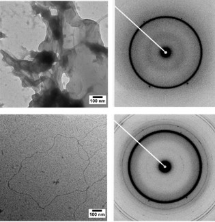
SAXS analysis revealed that these areas of high MT density had spacings similar to that of MT bundles found in the axon initial segment (Fig. 2a-g). Additionally,  electron microscopy measurements not only confirmed these spacings but also revealed bundle geometries strikingly similar to the geometries found within the axon initial segment (Fig. 2h-j). The research team uncovered that this Tau-mediated interaction between MTs is a balance between the polymeric resistance to inter-digitation and biologically-encoded weak charge-charge attractions between Tau on opposing MTs.  Previous cell free reconstitutions had not observed MT bundles due to the use of paclitaxel, a MT-stabilizing agent. Only through an aggregate of weak, Tau-mediated interactions along  MTs of sufficient length will MT bundles manifest, a finding that was bolstered by a later study that showed that decreasing paclitaxel concentrations lead to longer MTs and Tau-induced MT bundles [3].

Figure 2

Figure 2. SAXS and TEM show that Tau-assembled MTs in active bundles at 37˚ C in 2 mM GTP recapitulate key in vivo features in neurons and other cells. a, Azimuthally-averaged SAXS data show that all six wild-type Tau isoforms induce MT bundles, with Bragg peak positions consistent with hexagonal lattices (top six profiles), as opposed to just microtubule form factor for no Tau (bottom profile). b-e, Line-shape analysis of SAXS data [resultant fits in red on (a)] yields the ensemble-averaged microtubule inner radius <rin> (b), hexagonal lattice parameter aH (c), wall-to-wall distance Dw-w (d), and Dw-w normalized by the calculated Tau projection domain radius of gyration, RGPD (e). f, g, Dw-w and Dw-w/RGPD as a function of Tau net charge (QTau) shows a monotonic decrease in Dw-w and a nearly constant Dw-w/RGPD ≈ 8-11, respectively. The latter is especially surprisingly, as the expected Dw-w/RGPD should be ~ 4. h, Electron microscopy of microtubules assembled with Tau (Φ3RM=1/20) at low magnification show distinct bundled domains, demonstrating phase separation. i, Domains of hexagonally-ordered arrays of microtubules (identified in white outlines, Φ3RL=1/20) with vacancies likely resulting from the suppressed (but still occurring) dynamic instability. j, Linear bundles of microtubules (Φ3RL=1/20), a result of extensive vacancy introduction and mimicking string-like microtubule bundles in the axon initial segment. In (i) and (j) the staining process exaggerates the microtubule wall thickness. Scale bars, 1 µm (h) and 500 nm [(i) and (j)].   Adapted from [2].


The intrinsic disorder of Tau on MTs allows for a fascinating blend of both biopolymer and protein characteristics; not only can the N-terminal tail of Tau transition to a polyelectrolyte brush on MT surfaces as a function of Tau isoform and coverage density, but attractions between charged amino acids on opposing N-terminal tails of Tau allow for complex MT bundle geometries.

Here, the researchers demonstrate the power of the SAXS-osmotic pressure technique in elucidating the distinct conformations of the protruding N-terminal tail of Tau (namely, the mushroom and brush state at low and high coverage, respectively), fundamentally altering the force between MTs. The paper reported the discovery that the brush conformation of Tau is crucial in preventing aggregation of microtubules, which otherwise would result in the loss of their function as molecular rails in healthy neurons.

They further reconcile conflicting in vivo and cell free studies by revealing that Tau does, in fact, induce MT bundles in dissipative physiological conditions (i.e. in the absence of the MT stabilizing agent paclitaxel and under GTP hydrolyzing conditions). The key insight is that only an aggregate of this weak, Tau-mediated interactions along long MTs (such as those found within the axon initial segment) will recapitulate microtubule bundles found in the axon. This unique interaction is contingent on the intrinsic disorder of Tau.

The functionalization of these protein nanotubes by Tau, a polyampholyte, is inherently absent in charged polymers containing all positive or all negative charges (e.g. like DNA, which only contains negative phosphate groups). These novel, emerging properties gives insight for the design of biologically-inspired materials with multiple interaction motifs of opposite charge.

Acknowledgements

Research primarily supported by the US Department of Energy (DOE), Office of Science, Basic Energy Sciences (BES) under award number DE-FG02-06ER46314 (self-assembly and force measurements in filamentous protein systems), the US National Science Foundation (NSF) under award number DMR-1401784 (protein phase behavior), and the US National Institutes of Health under award numbers R01-NS13560 and R01-NS35010 (tubulin purification and protein Tau isoform purification from plasmid preparations). U.R. acknowledges support from the Israel Science Foundation (Grant 351/08). M.C.C. was supported by NRF-2014R1A1A2A16055715, 2014M2B2A4030706 and APCTP. The x-ray diffraction work was carried out at the Stanford Synchrotron Radiation Lightsource, a Directorate of SLAC National Accelerator Laboratory and an Office of Science User Facility operated for the US DOE Office of Science by Stanford University. The SSRL Structural Molecular Biology Program which supports BL4-2 is funded by the DOE Biological and Environmental Research and the NIH National Institute of General Medical Science (P41GM103393)

Primary Citation(s): 
P. J. Chung, C. Song, J. Deek, H. P. Miller, Y. Li, M. C. Choi, L. Wilson, S. C. Feinstein and C. R. Safinya, "Tau Mediates Microtubule Bundle Architectures Mimicking Fascicles of Microtubules Found in the Axon Initial Segment", Nat. Commun. 7, 12278 (2016), DOI: 10.1038/ncomms12278. P. J. Chung, M. C. Choi, H. P. Miller, H. E. Feinstein, U. Raviv, Y. Li, L. Wilson, S. C. Feinstein and C. R. Safinya, "Direct Force Measurements Reveal that Protein Tau Confers Short-range Attractions and Isoform-dependent Steric Stabilization to Microtubules", Proc. Natl. Acad. Sci. USA 112, E6416 (2015), DOI: 10.1073/pnas.1513172112.
References: 

[1] P. J. Chung, M. C. Choi, H. P. Miller, H. E. Feinstein, U. Raviv, Y. Li, L. Wilson, S. C. Feinstein and C. R. Safinya, "Direct Force Measurements Reveal that Protein Tau Confers Short-range Attractions and Isoform-dependent Steric Stabilization to Microtubules", Proc. Natl. Acad. Sci. USA 112, E6416 (2015), DOI: 10.1073/pnas.1513172112.

[2] P. J. Chung, C. Song, J. Deek, H. P. Miller, Y. Li, M. C. Choi, L. Wilson, S. C. Feinstein and C. R. Safinya, "Tau Mediates Microtubule Bundle Architectures Mimicking Fascicles of Microtubules Found in the Axon Initial Segment", Nat. Commun. 7, 12278 (2016), DOI: 10.1038/ncomms12278.

[3] M. C. Choi, P. J. Chung, C. Song, H. P. Miller, E. Kiris, Y. Li, L. Wilson, S. C. Feinstein and C. R. Safinya, "Paclitaxel Suppresses Tau-mediated Microtubule Bundling in a Concentration-dependent Manner", Biochim. Biophys. Acta 1861, 3456 (2017), DOI: 10.1016/j.bbagen.2016.09.011.

Beamlines Used:

Topic:

Structural Mechanisms of Histone Recognition by Histone Chaperones

February 2018

Chromatin is the complex of DNA and proteins that comprises the physiological form of the genome. Non-covalent interactions between DNA and histone proteins are necessary to compact large eukaryotic genomes into relatively small cell nuclei. The nucleosome is the fundamental repeating unit of chromatin, and is composed of 147bp of DNA wrapped around an octamer of histone proteins: 2 copies of each H2A, H2B, H3 and H4.

Assembly of nucleosomes in the cell requires the coordinated effort of many proteins including ATP-dependent chromatin remodeling enzymes and ATP-independent histone chaperone proteins. Histone chaperones are a large class of proteins responsible for binding the highly basic histone proteins, shielding them from non-specific interactions, facilitating nuclear import of histones, and finally depositing histones onto DNA to form nucleosomes. Despite performing many overlapping functions, histone chaperone proteins are highly structurally divergent. However, nearly all histone chaperones contain highly charged intrinsically disordered regions (IDRs)1. In many cases truncation of these conserved regions results in loss of histone affinity and deposition functions.

Nucleoplasmin (Npm) is a highly conserved H2A/H2B-specific histone chaperone expressed during the early stages of vertebrate development. Npm is tasked with storing large amounts of H2A/H2B dimers in the oocyte (an immature cell in the ovary), and releasing them upon fertilization to keep up with the rapidly dividing embryo. Structurally, Npm contains a pentameric N-terminal Core domain and an intrinsically disordered C-terminal Tail domain composed of alternating acidic and basic stretches (Figure 1). Previous mass-spectrometry analysis showed that Npm is heavily modified mainly along the Tail domain during development, and that these modifications alter its histone affinity and deposition rates2. Complete removal of this IDR indicated that it is necessary for histone binding, however smaller truncations of the C-terminus resulted in increased histone affinity and deposition rates. This suggested that this region acts as both a major site of histone interaction and a regulator of Npm function.

Npm fig 1

Figure 1: Domain organization of the histone chaperone Npm.  The N-terminal Core domain (residues 16-118) is ordered and forms a stable homopentamer.  The C-terminal Tail domain (residues 119-195) is disordered. Disorder prediction (DISOPRED3 score) shown below. The Tail domain contains the larges acidic stretch (A2) that directly engages H2A/H2B dimers, as well as a basic nuclear localization sequence and basic C-terminus that shield A2 and negatively regulate the function of Npm.


A study led by Christopher Warren and Dr. David Shechter at the Albert Einstein College of Medicine utilized NMR and biochemical assays to show that the intrinsically disordered Npm Tail domain negatively regulates histone binding by dynamic intramolecular shielding of a key acidic stretch (A2)3. Paramagnetic Relaxation Enhancement NMR (PRE-NMR) was used to gain structural information on the monomeric Tail domain alone and in complex with H2A/H2B. A stable pentameric complex of Npm bound to five H2A/H2B dimers was able to be formed by removal of this C-terminal autoregulatory region, though this complex was far too large for NMR structural analysis and resisted all crystallization attempts.

In a collaboration with Dr. Tsutomu Matsui, SSRL, size-exclusion chromatography coupled SAXS (SEC-SAXS) data were measured at BL4-2 for this complex (Figure 2). The SEC-SAXS data confirmed that pentameric complex was stable and without significant flexibility. The oblate, star shaped SAXS envelope calculated from these data indicated that the H2A/H2B dimers rest on the upper portion of the lateral face of the Npm pentamer. Using the relative positioning of the Tail domain and H2A/H2B obtained by PRE-NMR, Dr. Matsui was able to build novel models of the pentameric complex that satisfied structural restraints from both NMR and SAXS data. These NMR-restrained SAXS hybrid models provide the highest resolution insight to date on the architecture of a complex that is critical setting up the chromatin landscape in the earliest stages of embryonic development. These models also help to partially explain the vital role that Npm plays in histone storage and deposition processes during vertebrate development. Future collaborative studies will target the structural basis for understanding the roles of many histone chaperones in both normal and cancerous cells.

Npm fig 2

Figure 2: SAXS analysis of Npm Core+A2 truncation (1-145) bound to five H2A/H2B dimers. Left: small angle x-ray scattering curve of the complex (purple dots). Simulated SAXS curve from the best scoring structural model shown as a black line. Right: SAXS envelope of the complex (pink) with the best scoring structural model inside. Positioning of H2A/H2B dimers by NMR and SAXS structural restraints.


Primary Citation(s): 
C. Warren, T. Matsui, J. M. Karp, T. Onikubo, S. Cahill, M. Brenowitz, D. Cowburn, M. Girvin, D. Shechter, "Dynamic Intramolecular Regulation of the Histone Chaperone Nucleoplasmin Controls Histone Binding and Release", Nat. Commun. 8, 2215 (2017) doi:10.1038/s41467-017-02308-3.
References: 
  1. C. Warren and D. Shechter, "Fly Fishing for Histones: Catch and Release by Histone Chaperone Intrinsically Disordered Regions and Acidic Stretches", J. Mol. Biol. 429, 2401 (2017) doi: 10.1016/j.jmb.2017.06.005.
  2. T. Onikubo, J. J. Nicklay, L. Xing, C. Warren, B. Anson, W.-L. Wang, E. S. Burgos, S. E. Ruff, J. Shabanowitz, R. H. Cheng, D. F. Hunt, D. Shechter, "Developmentally Regulated Post-translational Modification of Nucleoplasmin Controls Histone Sequestration and Deposition", Cell Rep. 10, 1735 (2015) doi:10.1016/j.celrep.2015.02.038.
  3. C. Warren, T. Matsui, J. M. Karp, T. Okinubo, S. Cahill, M. Brenowitz, D. Cowburn, M. Girvin, D. Shechter, "Dynamic Intramolecular Regulation of the Histone Chaperone Nucleoplasmin Controls Histone Binding and Release", Nat. Commun. 8, 2215 (2017) doi:10.1038/s41467-017-02308-3.

Beamlines Used:

Topic:

Subscribe to RSS - BL4-2