Ecological and political realities have moved discussions of, and advances in, fuel cell technology into mainstream public awareness. Electrocatalysis, the science of modifying the overall rates of electrochemical reactions so that selectivity, yield and efficiency are maximized, is the work from which those advances spring. Studies in electrocatalysis have resulted in highly selective multicomponent gas mixture sensors, human blood component sensors, new electrocatalysts for oxidation/reduction of inorganic and organic pollutants in air and water, as well as better electrocatalysts for the fuel cell conversion of renewable and fossil fuels to electrical work. Studies of the mechanisms by which these catalysts operate have been advanced through development of in-situ surface x-ray scattering (SXS) techniques. SXS capabilities at SSRL were recently used to investigate the interface structure of an ultrathin COad (adsorbed carbon monoxide) overlayer on platinum. This work has elevated the macroscopic description of the COad state at the solid-liquid interface to a microscopic level and enabled the relation between the reactivity and the interfacial structure of COad/Pt to be understood.

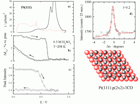
p(2 x 2)-3CO structure.
Of the various systems studied, CO oxidation [COad + OHad = CO2 + H+ + e-] on Pt and Pt-bimetallic surfaces occupies a special position in surface electrochemistry. Carbon monoxide is the simplest C1 molecule that can be electrochemically oxidized in a low temperature fuel cell at a reasonable (although not necessarily practical) potential. It thus serves as an important model "fuel" for fundamental studies of C1 electrocatalysis. For over a decade now the ability to characterize atomic/molecular spatial structures and to monitor changes in the local symmetry of surface atoms in-situ under reaction conditions has played an important part in our understanding of surface electrochemistry at metal-based interfaces [1,2]. This progress has been influenced greatly by the technique of in-situ surface x-ray scattering (SXS) which, in combination with infrared spectroscopy (IR) and electrochemical methods, has been used to find interrelationships between the microscopic surface structures of fcc metals (Pt, Ir, Pd, Au, Ag, Cu) and the macroscopic kinetic rates of the reactions. Two forms of COad species can be distinguished thermochemically on Pt(111) in an electrochemical environment [3]: (i) COad with a low heat of adsorption is characterized as the "weakly adsorbed" state, and (ii) COad with a relatively high enthalpy of adsorption is characterized as the "strongly adsorbed" state. In our recent studies, the macroscopic description of the COad state at the solid-liquid interface has been elevated to a microscopic level, which enables the relation between the reactivity and the interfacial structure of COad on Pt(111) to be understood.
Representative SXS results along with CO oxidation current in 0.05 M H2SO4 are summarized in Figure 1. Direct information regarding the induced relaxation of surface Pt atoms by the adsorption of CO are obtained by analyzing and modeling crystal truncation rod (CTR) data [4] (not shown). The potential dependence of the Pt(111) surface relaxation, denoted as x-ray voltammetry (XRV) induced by COad adsorption is represented by the results in Figure 1b. In COad-free solution at 0.05 V the top Pt atomic layer expands ca. 2 % (0.05 Å) of the lattice spacing away from the second atomic layer when the adsorbed hydrogen (Had) reaches its maximum coverage (0.66 ML).
Following the adsorption of CO at 0.05 V, the Pt surface expansion is even larger ca. 4 %. The difference in relaxation of the Pt(111) surface covered with Had ad probably arises from the difference in the adsorbate-metal bonding, the Pt(111)-COadad interaction. At 0.05 V, no change in the relaxation of the Pt surface atoms was observed after replacement of CO from solution with nitrogen, indicating that CO ad is indeed irreversibly adsorbed on the Pt(111) surface. Upon sweeping the potential positively from 0.05 V, the oxidation of COad in the so-called pre-oxidation potential region (0.3 < E < 0.6 V) is mirrored with a small contraction of the Pt surface layer. Above ca. 0.6 V, the top layer expansion is reduced significantly, contracting above 0.7 V to the unrelaxed state that the Pt(111) surface has in the absence of CO ad.

Direct information regarding the COad structure on Pt(111) was obtained by searching in the surface plane of reciprocal space for diffraction peaks characteristic of an ordered adlayer. At 0.05 V, a diffraction pattern consistent with a p(2 x 2) symmetry was observed between
0.05 < E < 0.6 V. A rocking scan through the (½, ½, 0.2) position together with the derived structural model, which consists of three CO molecules per p(2 x 2) unit cell, is shown schematically in Figure 1d. From the width of this peak and from the result of similar fits to other p(2 x 2) reflections a coherent domain size in the range of 80-120 Å for the CO adlayer was deduced. Upon the reversal of the electrode potential at ca. 0.6 V, the p(2 x 2)-3CO structure is not re-formed, Figure 1c, confirming that the structure is coverage-dependent and not just potential-dependent.
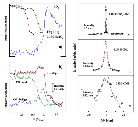
With a constant overpressure of CO in the x-ray cell, the SXS experiments revealed a reversible loss and re-formation of the p(2 x 2)-3CO structure, with the p(2 x 2)-3CO structure re-forming as the potential was slowly (1 mV/s) swept below 0.2 V, Figure 2a. IR has provided a valuable complement to the structural information obtained from SXS [3]. As shown in Figure 2b, the spectra for CO ad on Pt(111) in CO-saturated 0.1 M HClO 4 have three characteristic Pt-CO stretching frequencies, the atop CO near 2070 cm -1, the multi-coordinated COad near 1780 cm -1, and the bridge CO ad near 1840 cm -1. Figure 2 shows that oxidation of the COad in the three-fold hollow sites and relaxation of the remaining COad into bridge sites and atop sites is accompanied with both the decrease in the Bragg peak intensity for the p(2 x 2)-3CO structure and CO2 formation. Therefore, the macroscopic characterization of a weakly adsorbed state is linked microscopically to a saturated CO adlayer consisting of three COad molecules per p(2 x 2) unit cell located in a-top and three-fold hollow sites of the Pt(111) surface. Figure 2 shows that oxidation of the COad in the three-fold hollow sites and relaxation of the remaining COad into bridge sites and atop sites disrupts the long-range ordering in the remaining adlayer, as the Bragg peak intensity for the p(2 x 2)-3CO structure decreases rapidly in this potential region. The "relaxed" COad adlayer (characterized microscopically) with substantial alternation in binding site geometry from predominantly three-fold hollow to bridge sites can be linked macroscopically to the strongly adsorbed state of COad that is oxidized in the ignition potential region. It is also worth mentioning that besides tuning the fine balance between atop and multifold coordinated COad, the domain size of the COad structure is significantly affected by the nature of anions. For example, the domain size (Figures 2d-2f ) and stability (not shown) of the p(2 x 2)-3CO structure increases from KOH (ca. 30 Å), to HClO4 (ca. 140 Å) to HClO4 + Br- (ca.350 Å); i.e., the less active the surface is towards COad oxidation, the larger the ordered domains of the p(2 x 2)-3 CO structure. As discussed in reference [3], a self-consistent explanation for this result is that both the stability and domain size of the ordered COad adlayer are determined by the competition between OHad and spectator anions for the defect/step sites on the Pt(111) surface.
Acknowledgment
This work was supported by the Assistant Secretary for Conservation and Renewable Energy, Office of Transportation Technologies, Electric and Hybrid Propulsion Division of the U.S. Department of Energy under Contract No. DE-AC03-76SF00098. Research was carried out in part at SSRL which is funded by the Division of Chemical Sciences(DSC), U.S. DOE. CAL acknowledges the support of an EPSRC Advanced Research Fellowship.
- C. A. Lucas and N. M. Markovi, in 'Encyclopedia of Electrochemistry', Volume 2 (Wiley-VCH, 2003) Chapter 4.
- N. M. Markovic and P.N. Ross, Surf. Sci. Rep., 45 (2002) 121-229.
- N.M. Markovic, C.A. Lucas, A. Rodes, V. Stamenkovic, P.N. Ross, Surf. Sci., 499 (2002), L149-L158.
- C. A. Lucas, N.M. Markovi, P.N. Ross, Surf. Sci., 425 (1999) L381-L386.
N. M. Markovic, C. A. Lucas, A. Rodes, V. Stamenkovic, and P. N. Ross "Surface Electrochemistry of CO on Pt(111): Anion Effects", Surf. Sci. 499, L149 (2002)

