The adaptive immune response enables the vertebrate immune system to recognize and respond to specific pathogens, with immunological memory allowing a stronger response upon subsequent re-exposure to a pathogen. Adaptive immunity relies on the capacity of immune cells to distinguish between the body's own cells and foreign invaders. αβ T cell receptors (TCRs) recognize antigenic peptides in complex with major histocompatibility complex proteins (MHC) as the central event in the cellular adaptive immune response. Despite undergoing an extensive education process in the thymus, mature T cells exhibit a high frequency of crossreactivity, or alloreactivity, toward foreign peptide-MHC to which they have not previously been exposed. Alloreactivity indicates an inherent ability of the TCR to crossreact with a broad range of self and foreign peptide-MHC ligands, which could be beneficial for immune surveillance of a universe of potential pathogens. However, it presents a major clinical problem for organ transplantation in that genetically mismatched tissue can be rejected in a graft-host alloresponse.
The molecular basis of alloreactivity remains poorly understood, despite the fact that the general structural principles of TCR/pMHC interactions have been defined in approximately 14 cocrystal structures (Rudolph et al., 2006). More broadly, receptor-ligand crossreactivity has been observed across many systems and has generally been attributed to "molecular mimicry." However, structural evidence for molecular mimicry, in the form of complexes between one receptor and multiple similar, but distinct ligands, remains elusive for most systems.

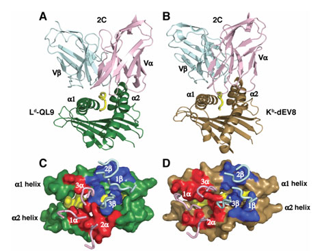
The ability of a TCR to directly recognize foreign (allogeneic) MHC molecules underlies T cell-mediated rejection in patients receiving allogeneic organ transplants. In order to better understand the differences in recognition of self versus foreign peptide-MHC complexes, we crystallized and solved the structure of the 2C TCR bound to a foreign MHC, collecting data at SSRL beamline 11-1. We compared our TCR/foreign MHC structure to the previously published structure of the same TCR bound to a self MHC (Garcia et al., 1998), revealing that instead of mimicking the interactions formed with a self MHC, a single TCR adopts a completely different strategy to recognize a foreign MHC (Figure 1). This is surprising, since the self and foreign MHC share 80% sequence identity in the helices presented to the TCR. Very few conserved interactions are found in the allogeneic and syngeneic complexes, and these occur via largely unique binding chemistries.
In addition, there does not appear to be a focus on the polymorphic MHC residues presented to the TCR. Thus, while sequence conservation between the self and foreign ligands might have suggested similar recognition strategies, the interatomic contacts highlight a vastly different mode of recognition. In addition to the co-complex structure described, we also crystallized and solved the structure of an engineered, high-affinity variant of the 2C TCR bound to the same foreign MHC. In this structure, the data for which were also collected on beamline 11-1 at SSRL, the "wild-type" recognition footprint persists despite modified interactions with the peptide, indicating that differences in the antigenic peptide are not necessarily sufficient to alter TCR recognition.
Thus, we show that a single TCR recognizes two globally similar, but distinct ligands by divergent mechanisms, indicating that receptor-ligand crossreactivity can occur in the absence of molecular mimicry.
This work was supported by NIH grants AI 48540 (K.C.G.) and GM55767 (D.M.K.), a National Science Foundation predoctoral fellowship (L.C.), the Keck Foundation, and the Howard Hughes Medical Institute.
- Garcia, K.C., Degano, M., Pease, L.R., Huang, M., Peterson, P.A., Teyton, L., and Wilson, I.A. (1998). Structural basis of plasticity in T cell receptor recognition of a self peptide-MHC antigen. Science (New York, NY 279, 1166-1172.
- Rudolph, M.G., Stanfield, R.L., and Wilson, I.A. (2006). How TCRs bind MHCs, peptides, and coreceptors. Annual review of immunology 24, 419-466.
Colf, L.A., Bankovich, A.J., Hanick, N.A., Bowerman, N.A., Jones, L.L., Kranz, D.M., and Garcia, K.C. (2007). How a single T cell receptor recognizes both self and foreign MHC. Cell 129, 135-146.


