

Figure 1. Illustration of the nanoscopic study of a BiNiO3 particle under high pressure.
BiNiO3 is of great fundamental interest and practical importance thanks to its colossal negative thermal expansion behavior. This unusual property may be used to fabricate composites with zero or other controlled thermal expansion values, which have a range of potential engineering, photonic, electronic, and structural applications, and thus has attracted lots of research attention. The same low-density to high-density phase transition accompanying an increase in temperature can also be induced by changing the external pressure which can be well controlled using high-pressure vessels such as diamond anvil cells. Numerous studies on BiNiO3 have been carried out using bulk analysis tools such as x-ray diffraction and x-ray spectroscopy. However, the detailed evolution of the phase transition/propagation, which is the key to a deeper understanding of the mechanism behind the measured bulk properties, remains unknown.
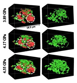
Figure 2. Low-pressure-phase (red) and high-pressure-phase (green) regions of the BiNiO3 particle as a function of the external pressure.
Scientists from Stanford Synchrotron Lightsource (Yijin Liu), the Center for High Pressure Science and Technology Advanced Research in Shanghai, China and Carnegie Institute of Washington (Wenge Yang and Junyue Wang), Tokyo Institute of Technology (Masaki Azuma), and Stanford University and SLAC (Wendy L. Mao) designed an experiment to study the intermediate states of the phase transition by performing nanoscopy
on a BiNiO3 particle at elevated pressure as high as a few gigapascals. This novel experimental scheme, illustrated in Fig. 1, allows the researchers to investigate the interesting reversible phase transition by probing the particle under the required condition for the transition. A clear five-dimensional (X, Y, Z, x-ray energy, pressure) visualization of this process, which shows the change of the chemically resolved three-dimensional morphology as a function of external pressure, was obtained by the researchers.
The phase front propagation (see Fig. 2) was resolved and visualized for the first time and with unprecedented spatial resolution using state-of-the-art nanoscale synchrotron x-ray tomography at SSRL’s Beam Line 6-2C. The result provides important implications for the evolution of the pressure-induced phase transition. The capability of resolving and visualizing the nucleation and growth of the new phase opens vast scientific opportunities in the high-pressure research field.
Y. Liu, J. Wang, M. Azuma, W. L. Mao, and W. Yang, “Five-dimensional Visualization of Phase Transition in BiNiO3 under High Pressure”, Appl. Phys. Lett. 104, 043108 (2014), DOI: 10.1063/1.4863229.
