
Ultrafast x-ray scattering and spectroscopic measurements have captured snapshots of the atomic-scale mechanisms underlying how a class of superionic nanocrystals transform. Superionic materials are multi-component solids with simultaneous characteristics of both a solid and a liquid: Above a critical temperature associated with a structural phase transition, one atomic species in the material exhibits liquid-like ionic conductivities and dynamic disorder within the rigid crystalline structure of the other. Applications such as electrochemical storage materials and resistive switching devices follow from this abrupt change in ionic mobility, but the atomistic pathways associated with this phase transition and the related functional properties that emerge at the nanoscale are largely unknown and unexplored. These measurements, carried out at the Stanford Synchrotron Radiation Lightsource (SSRL), the Advanced Light Source (ALS) and the Advanced Photon Source (APS), capture for the first time the atomic-level dynamics of a nanocrystal as it transforms – and do so with femtosecond temporal resolution.
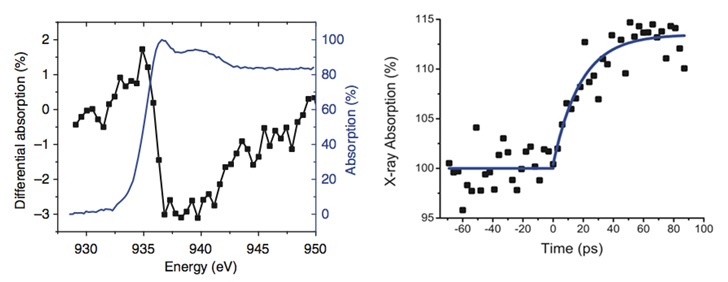
Measurements probed the dynamics of copper sulfide (Cu2S) nanodiscs (10 nm diameter) deposited on silicon nitride membranes. X-ray spectroscopic measurements were made using femtosecond soft x-ray pulses generated at the ALS. Measurements probed photo-induced changes in the local bonding geometry at the Cu L-edge in transmission with elemental specificity. Additional temperature-dependent measurements of the spectroscopic changes associated with the phase transition were carried out as a function of temperature at SSRL Beam Line 13-3 in total electron yield. Hard x-ray scattering measurements were carried out at SSRL and APS in order to characterize the long-range structural changes associated with the phase transition. Fig. 2 shows optically-induced changes in the nanocrystal scattering pattern associated with the transition between the low-temperature and high-temperature structural phases recorded at SSRL Beam Line 2-3. Fig. 3 shows the transient differential energy spectrum at t=100 ps and the time-scale for the observed redshift in the spectrum, measured with 200-femtosecond time resolution using short-pulse x-rays at ALS's Beam Line 6.0.2. Through detailed modeling of the structural changes that occur across the phase transition temperature, we associate this redshift with the onset of superionicity.
Together, these measurements show that the intrinsic time for the transformation is governed by the ionic hopping time in these systems, identifying a transition state in which diffusional motion of the ions leads to the formation of the superionic crystallographic phase. They further show that the transition can occur on picosecond time-scales, identifying the possibility of nanoscale superionic-based switching devices controllable by light and operating with speeds orders of magnitude faster than previously demonstrated.
Research supported by the Department of Energy, Basic Energy Sciences, Materials Sciences and Engineering Division.
T. A. Miller, J. S. Wittenberg, H. Wen, S. Conner, Y. Cui, A. M. Lindenberg, "The Mechanism of Ultrafast Structural Switching in Superionic Copper (I) Sulphide Nanocrystals", Nature Communications 4,1369 (2013).
