Botulinum neurotoxin (BoNT), the most potent toxin known, induces a potentially fatal paralytic condition known as "botulism". Botulism can occur when toxin-producing bacteria infect wounds (wound botulism) or the intestinal tract (infant/intestinal botulism), or following the ingestion of contaminated food in which toxin has been produced (food-borne botulism). In the USA, infant botulism represents the most common manifestation of the disease, where its prevalence has led to speculation of a link to sudden infant death syndrome. BoNTs are subdivided into seven distinct serotypes (types A through G), and an increasingly large number of subtypes continue to be identified within each serotype, highlighting the need to produce broad-spectrum therapeutics. BoNT variants are an important biochemical set of tools for understanding nerve function, and important therapeutic agents in current clinical use to provide relief to patients with a wide spectrum of neurological disorders.
Recently, the Stevens Laboratory at The Scripps Research Institute, in collaboration with the Marks laboratory at UCSF and the Chapman and Johnson laboratories at the University of Wisconsin, Madison, completed structural studies on the structures of botulinum toxin in complex with the neuronal cell surface receptor synaptotagmin II (Syt-II) recognition domain (1) and botulinum toxin with two different neutralizing monoclonal antibodies (2). To compliment the structural work, biochemical, mutagenesis, and cellular assay experiments were also completed. The interdisciplinary research projects provide insight into the atomic details on the intoxication process, and ways that antibodies can neutralize the effects. These structures open the possibility of developing improved broad-spectrum therapeutics, including antibodies, small molecule drugs and vaccines against the toxin.

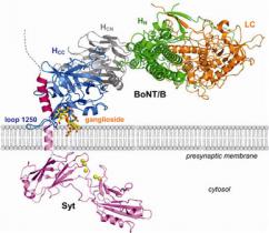
The first structural study is that of the BoNT/B-Syt-II complex at 2.6 Å resolution (1). This work reveals a possible structural basis to help understand the remarkable neuron specificity and extreme potency of BoNTs. Decades ago, a "double receptor" model was proposed in which BoNTs recognize nerve terminals via interactions with both gangliosides and protein receptors that mediate their cell entry (3). Among the seven BoNTs, the putative receptors for BoNT/A, /B (4-5) and /G have been identified, yet the molecular details that govern recognition remained unclear. The structure of the complex reveals that Syt-II adopts a helical conformation on binding to a hydrophobic groove within the binding domain of BoNT/B. This is further validated by mutagenesis of residues on Syt-II in this region, carried out as part of our studies, which is observed to negatively affect BoNT/B binding. In addition, our molecular docking studies using the ganglioside GT1b indicate that its binding site is more extended than previously proposed, and possibly forms contacts with both BoNT/B and Syt. The structure of the BoNT/B-Syt-II complex with modeled ganglioside discloses an enlightening molecular snapshot of BoNT/B while anchored to the presynaptic membrane (Fig. 1). When both ganglioside and Syt-II binding are presented, the C-terminal trefoil subdomain (HCC) of BoNT/B appears to be locked onto the cell surface at one end by the two anchor points. Thus, our study presents a structural basis for the long speculated "double receptor" hypothesis, and also provides valuable information for the development of inhibitors that may block binding of toxins to cell surface receptors. Most importantly, it suggests that the development of inhibitors that disrupt the synergetic effects brought on by the double receptor binding during complex formation should be a therapeutic with exceptional potency, given the amplified effect of blocking both receptor binding sites simultaneously. Additionally, the knowledge of specific interaction of BoNT with its receptors provides a rational basis for designing an engineered BoNT that targets different cell types other than motor neurons, expanding its use to a wider array of clinical applications. An adjoining elegant article in the same issue of Nature by the Brunger laboratory at Stanford provides additional insight into this incredible set of interactions that help to define multivalent specificity and affinity (6).

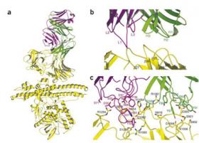
In the second publication, we determined the X-ray co-crystal structures of wild-type and cross-reactive antibodies (AR2 and CR1) complexed to BoNT/A1 at resolutions up to 2.6 Å (2). Both Fabs bind to identical regions on the BoNT/A1 binding domain, at the interface between the N-terminal lectin subdomain (HCN) and the C-terminal trefoil subdomain (HCC) (Fig. 2). A combination of hydrophilic and hydrophobic interactions is responsible for forming the complex. However, AR2 scFv (single chain variable fragment) binds the BoNT/A1 subtype with high affinity (136 pM) and the BoNT/A2 subtype with low affinity (109 nM). The engineered scFv CR1 displays 1,250-fold increased affinity for BoNT/A2 (87 pM), while maintaining high-affinity binding to BoNT/A1 (115 pM). Structural analysis revealed that the increased affinity of CR1 for BoNT/A2 results from the amino acid differences between the antibodies in the H1 loops: S30K, D31Y and H32D (Fig. 2c). Given the amino acid variability observed among seven serotypes and hundreds of subtypes of BoNT, our structures of the complex provide a powerful basis for protein engineering that can be used to fine tune antibody specificity and broaden cross-activity.
These works were supported by a grant from the Pacific Southwest Regional Center of Excellence (R.C.S. and E.A.J.). Portions of this research were carried out on beam lines 11-1 and 1-5 at the SSRL, a national user facility operated by Stanford University on behalf of the US Department of Energy, Office of Basic Energy Sciences. The SSRL Structural Molecular Biology Program is supported by the Department of Energy, Office of Biological and Environmental Research and by the National Institutes of Health, National Center for Research Resources, Biomedical Technology Program, and the National Institute of General Medical Sciences.
- Chai Q, Arndt JW, Dong M, Tepp WH, Johnson EA, Chapman ER, Stevens RC. Structural basis of cell surface receptor recognition by botulinum neurotoxin B. Nature 2006, 444, 1096-1100
- Garcia-Rodriguez C, Levy R, Arndt JW, Forsyth CM, Razai A, Lou J, Geren I, Stevens RC. Molecular evolution of antibody cross-reactivity for two subtypes of type A botulinum neurotoxin. Nature Biotechnol. 2007, 25, 107-116
- Montecucco, C. How do tetanus and botulinum toxins bind to neuronal membranes? Trends Biochem. Sci. 1986, 11, 315-317.
- Nishiki T, Tokuyama Y, Kamata Y, Nemoto Y, Yoshida A, Sato K, Sekiguchi M, Takahashi M, Kozaki S. The high-affinity binding of Clostridium botulinum type B neurotoxin to synaptotagmin II associated with gangliosides GT1b/GD1a. FEBS Lett. 1996, 378, 253-7.
- Dong M, Richards DA, Goodnough MC, Tepp WH, Johnson EA, Chapman ER. Synaptotagmins I and II mediate entry of botulinum neurotoxin B into cells. J. Cell. Biol. 2003, 162, 1293-303.
- Jin R, Rummel A, Binz T, Brunger AT. Botulinum neurotoxin B recognizes its protein receptor with high affinity and specificity. Nature 2006, 444, 1092-1095.
Chai Q, Arndt JW, Dong M, Tepp WH, Johnson EA, Chapman ER, Stevens RC. Structural basis of cell surface receptor recognition by botulinum neurotoxin B. Nature 2006, 444, 1096-1100
Garcia-Rodriguez C, Levy R, Arndt JW, Forsyth CM, Razai A, Lou J, Geren I, Stevens RC. Molecular evolution of antibody cross-reactivity for two subtypes of type A botulinum neurotoxin. Nature Biotechnol. 2007, 25, 107-116


