Membrane proteins are notoriously difficult to crystallize, and fiber-forming proteins were actually declared "uncrystallizable" by the eminent x-ray crystallographer Sir Lawrence Bragg. Supported by the facilities and staff at SSRL, a team of researchers has recently determined structures that solved both problems by defining the atomic structure of the membrane-associate protein pilin and its assembled Type IV pilus (T4P) fiber structure. T4P are filamentous organelles displayed on the surfaces of most Gram-negative bacteria 1. T4P are central to host colonization for many bacterial pathogens, mediating diverse and essential functions such as motility, adhesion, microcolony formation and uptake of DNA and specific filamentous phage. T4P are several microns in length and only 60-90 Å in diameter, and are comprised of thousands of copies of a single subunit, the pilin protein. Type IV pilins from different bacterial species share a common sequence of mostly hydrophobic amino acids in their N-terminus, as well as a pair of cysteines in their C-terminus, but differ substantially beyond these sites. Their prominent exposure on the bacterial surface and their key functions in virulence make T4P attractive targets for vaccines and therapeutics, the design of which would greatly benefit from their detailed molecular structures. Structural analyses of the Type IV pili have, however, proved to be extremely challenging. The presence of the hydrophobic N-terminal segment, which allows pilin to assemble and disassemble from the cell membranes, makes the pilin subunits insoluble without detergent. This problem has hindered efforts to crystallize the full-length proteins. Furthermore, the extremely thin and featureless pilus filaments reveal scant information on their helical symmetry, and thus resist classical helical image reconstruction approaches using electron micrograph (EM) images.
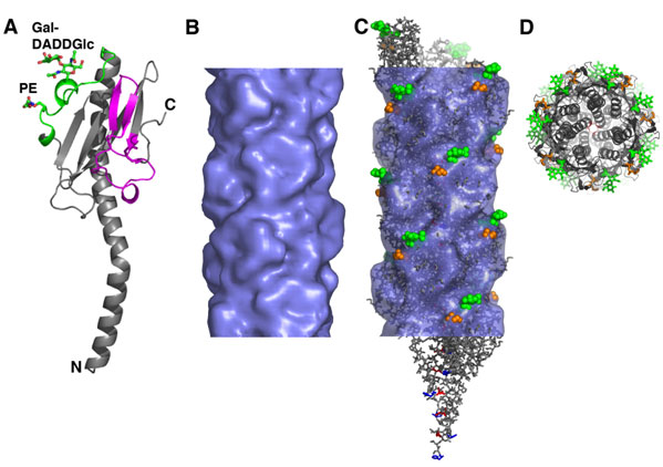
John Tainer and colleagues at the Scripps Research Institute have succeeded in solving the crystal structures of Type IV pilins from several important human pathogens using the macromolecular crystallography beamlines at SSRL (Beamlines 7-1, 9-1, 9-2 and 11-1). This success built directly upon the earlier efforts of Tainer group members Hans Parge and Andrew Arvai who made the original breakthrough pilin structure determination as aided enormously by the then new area detectors installed at SSRL according to John Tainer 2. Full length pilin structures were determined for Neisseria gonorrhoeae and Pseudomonas aeruginosa, and a truncated structure lacking the hydrophobic N-terminus was determi ned for Vibrio cholerae 2, 3. In a recently published article in Molecular Cell, this group report the 2.3 Å structure of N. gonorrhoeae (GC) pilin along with a "pseudo-atomic" structure of the pilus filament, solved by combined cryoEM reconstruction methods and computational docking of the pilin crystal structure 4. The N. gonorrhoeae Type IV pili are displayed peritrichously on the bacterial surface and mediate a multitude of functions in virulence, including immune escape. They share substantial sequence identity with the Type IV pili of N. meningitidis, which causes meningitis. The GC pilin crystal structure nicely extends the previous structure solved by the same group 2, and furthermore reveals the precise identity of two unusual post-translational modifications: an O-linked galactose (a1→3) diacetamidodideoxyglucose at Ser63 and a phosphoethanolamine at Ser68 (Fig. 1A). These post-translational modifications undergo phase variation and may also vary in structure from one generation of N. gonorrhoeae to the next, and have been proposed to function in immune escape. Another unusual feature of the pilin subunits is the "hypervariable loop", a segment between residues 128 and 141 that undergoes extreme sequence variation. This loop protrudes from the back of the pilin globular domain. The remainder of the GC pilin protein looks similar to other pilins: the hydrophobic N-terminus forms an extended a-helix, half of which is embedded in a b-sheet within the C-terminal globular domain.

Tainer and colleagues collaborated with Ed Egelman at the University of Virginia to determine a cryoEM reconstruction of the GC pilus filament using Egelman's Iterative Helical Real Space Reconstruction software 5. Although the filaments appear to be very smooth and featureless when examined by EM, they are in fact highly corrugated (Fig. 1B). The pilus structure was built by computationally fitting the pilin subunit structure into the cryoEM density and generating a filament using the symmetry operators determined for the reconstruction. In this pseudoatomic resolution pilus structure, deep grooves run between the subunits, and ridges formed by the post-translationally modified regions and the hypervariable loop protrude from the filament surface (Fig. 1C). The filaments are held together primarily by the N-terminal segments, which form a helical array in the core of the filament (Fig. 1D).
The N. gonorrhoeae Type IV pilus structure provides important insights into both pilus assembly and functions in pathogenesis. The filament can be viewed as three helical pilin polymers that twist around each other. These polymers are likely to be assembled simultaneously at the inner membrane of the bacteria, from a reservoir of pilin subunits that are anchored in the membrane via their hydrophobic N-termini. Importantly, the pilin and pilus structures provided the basis for a unified model for the assembly of these filaments that was recently supported by structures of the secretion superfamily ATPase 6, which were also done partly at SSRL and show the basis for the piston-like shifts to push T4P out by two helical turns. The grooves that run along the filament surface are positively-charged and represent an ideal, non-specific binding site for DNA, which can be brought into the cell by pilus retraction. And the locations of the variable post-translational modifications and hypervariable loop on protruding ridges on the filament would mask recognition of more conserved regions of the protein by protective antibodies. This structure is the first such structure determined for a Type IV pilus filament. The overall architecture of the filament is expected to be shared by all Type IV pili based on sequence and structure conservation for the pilin subunits. It is the regions of the subunit that vary, quite dramatically, among the pilins, that are exposed on the pilus surface and define its chemistry and hence its functions. This molecular understanding of a Type IV pilus filament reveals new targets for antibacterial agents that may have broad specificity.
This work was supported by NIH grants AI22160 (JAT, LC), EB001567 (EHE) and GM076503 (NV) and a fellowship from The Canadian Institutes of Health Research (LC).
- Craig, L., Pique, M. E. & Tainer, J. A. Type IV pilus structure and bacterial pathogenicity. Nat Rev Microbiol 2, 363-378 (2004).
- Parge, H. E. et al. Structure of the fibre-forming protein pilin at 2.6 A resolution. Nature 378, 32-8 (1995).
- Craig, L. et al. Type IV pilin structure and assembly: X-ray and EM analyses of Vibrio cholerae toxin-coregulated pilus and Pseudomonas aeruginosa PAK pilin. Molecular Cell 11, 1139-50 (2003).
- Craig, L. et al. Type IV pilus structure by cryo-electron microscopy and crystallography: implications for pilus assembly and functions. Molecular Cell 23, 651-662 (2006).
- Egelman, E. H. A robust algorithm for the reconstruction of helical filaments using single-particle methods. Ultramicroscopy 85, 225-34 (2000).
- Yamagata, A. & Tainer, J. A. Hexameric structures of the archaeal secretion ATPase GspE and implications for a universal secretion mechanism. Embo J 26, 878-90 (2007).
Craig, L. et al. Type IV Pilus Structure by Cryo-Electron Microscopy and Crystallography: Implications for Pilus Assembly and Functions. Molecular Cell 23, 651-662 (2006).


