With the ever-increasing demand to move away from fossil fuels and toward clean, renewable energy, dramatic improvements in energy storage devices are essential. Rechargeable lithium-sulfur (Li-S) batteries hold great potential for high-performance energy storage systems because they have a high theoretical specific energy, low cost, and are eco-friendly.1,2 However, a detailed understanding of the Li-S battery operating mechanism is vital to designing improvements for higher efficiency and capacity.
Until now, X-ray studies of Li-S electrodes have been done on batteries that were brought to a particular state of charge, allowed to relax, and then disassembled before measurement (an ex situ process). But further changes can occur within the battery during relaxation and disassembly, and such measurements may not necessarily give a true picture of battery chemistry and morphology. Additionally, the same region of a single battery cannot be followed throughout the charging and discharging cycle.

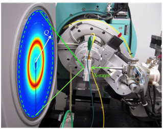
In this LDRD-supported project, scientists from SLAC and Stanford University have used a two-pronged approach to characterize Li-S batteries during normal battery operation. In operando X-ray Diffraction (XRD) (SSRL BL11-3) was used to characterize real-time changes in the active battery material’s crystal structure (Figure 1); while in operando X-ray imaging (SSRL BL6-2C) tracked nanometer-sized changes in the material’s morphology at 30 nm resolution. By using two complementary techniques during normal battery operation, a more complete picture of the battery mechanism was constructed. The results have provided new information different from previous ex situ studies. This highlights the importance of in operando studies and suggests possible strategies for improving cycle life.
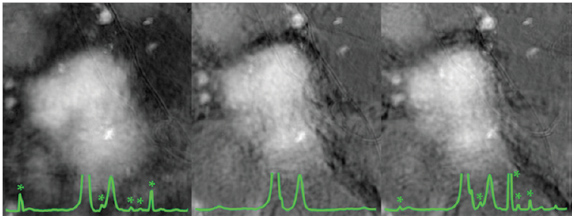
The in operando X-ray diffraction studies showed that during the discharge cycle crystalline sulfur interacted with lithium to form amorphous polysulfides. At the end of the discharge cycle crystalline Li2S did not form (Figure 2), which contradicts most published ex situ XRD studies (see for example Yuan et al.3). In fact, crystalline Li2S was only detected in a Li-S battery which was rested and disassembled. In operando X-ray imaging showed that polysulfides dissolved in the electrolyte, but they remained largely trapped within the electrode’s carbon matrix (Figure 2). This trapping will decrease fading of battery capacity due to loss of polysulfides to the electrolyte, which would dramatically reduce the lifetime of the battery.2 However, ex situ imaging of similar electrodes showed a complete absence of sulfur species on a partially discharged electrode. It is believed that during the disassembly of the battery, which includes thoroughly washing the electrolyte off the electrode, the dissolved polysulfides are freed. Thus, ex situ studies cannot accurately determine mechanisms for battery electrode failure.

In operando X-ray studies are essential to avoid misleading results and to understand more completely the changes occurring in active battery material during operation. Such studies not only characterize strengths and flaws of current battery technologies, but also can guide future electrode designs to either avoid or promote certain crystal structures and morphologies, in order to increase capacity in subsequent cycles and to prolong battery life. The in operando XRD and X-ray imaging work on Li-S has been published in the Journal of the American Chemical Society.
This work is supported by the Department of Energy, Laboratory Direct Research and Development funding, under contracts DE-AC02-76SF00515 (J.N., J.C.A., M.F.T.) and DE-AC02-76SF00515 (Y.C.). Y.Y. acknowledges support from a Stanford Graduate Fellowship, and A.J. acknowledges support from the Department of Defense (DoD) through the National Defense Science & Engineering Graduate Fellowship (NDSEG) Program. SSRL is supported by the Department of Energy, Office of Basic Energy Sciences.
- V. Etacheri, R. Marom, R. Elazari, G. Salitra, D. Aurbach, Challeges in the Development of Advanced Li-ion Batteries: a review. Energy & Environmental Science 4, 3243-3262 (2011).
- X. Ji, L. F. Nazar, Advances in Li-S Batteries. Journal of Materials Chemistry 20, 9821-9826 (2010).
- L. Yuan, X. Qiu, L. Chen, W. Zhu, New Insight into the Discharge Process of Sulfur Cathode by Electrochemical Impedance Spectroscopy. Journal of Power Sources 189, 127-132 (2009).
- H. Wang, Y. Yang, Y. Liang, J. T. Robinson, Y. Li, A. Jackson, Y. Cui, H. Dai, Graphene-Wrapped Sulfur Particles as a Rechargeable Lithium-Sulfur Battery Cathode Material with High Capacity and Cycling Stability. Nano Letters 11, 2644-2647 (2011).
J Nelson, S Misra, Y Yang, A Jackson, Y Liu, H Wang, H Dai, JC Andrews, Y Cui, MF Toney “In Operando X-ray diffraction and transmission X-ray microscopy of Lithium Sulfur Batteries” (2012) JACS 134, 6337-6343.


