Real-time measurement and control of the non-equilibrium properties of materials represents one of the 'grand challenges' in materials science and condensed matter physics. The ability to record snapshots of processes as they occur with atomic-scale spatial resolution and femtosecond temporal resolution extends these techniques to the level of atoms or electrons, with important applications to energy-related science and information storage technologies. With the advent of the LCLS, our ability to perform this kind of science will be dramatically enhanced. We have used a precursor to the LCLS, the Sub-Picosecond Pulse Source, to measure the first steps in the ablation process, extending the time-scales on which x-rays can be used to probe disordered materials by two orders of magnitude.

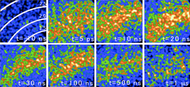
Under intense optical excitation, it is known from both ultrafast optical [1] and x-ray [2-5] studies that semiconductors transform to a disordered/liquid state on ultrafast time-scales, with large-amplitude atomic motions developing in hundreds of femtoseconds. However, the subsequent evolution of the liquid state has remained difficult to elucidate at atomic-scale resolution. Understanding of these processes forms the basis for the ablation process and laser-based materials processing. We have used femtosecond x-ray diffraction techniques to record the diffuse x-ray scattering pattern from InSb samples at excitation fluences near the ablation threshold of the material. Under these conditions, isochoric heating of the optically-formed liquid generates high temperature/high pressure conditions which relax into the liquid-vapor coexistence region of the phase diagram, leading to a rapid phase-explosion-driven boiling process within the disordered liquid. Figure 1 shows raw x-ray scattering data as collected by a CCD detector placed close to the sample, with x-rays incident at grazing incidence to the sample.
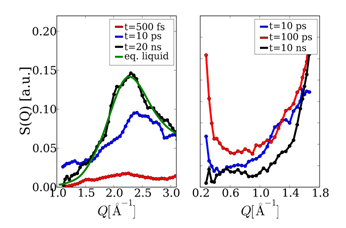
One observes the appearance of a diffuse scattering ring which narrows, shifts to lower Q, and eventually dissolves away as the liquid surface layer recrystallizes on hundreds of ns time-scales. In Figure 2, lineouts of these images are displayed at various times in both the wide angle and small angle scattering regimes. The development on ps time-scales of a transient divergence in the scattering at low Q is observed which can be associated with the formation of large amplitude density fluctuations in the structure of the liquid. By comparison to the structure factor for the equilibrium liquid (also shown) it is seen that for times up to 20 ns after excitation, the structure of the non-equilibrium liquid state is significantly different from the equilibrium liquid.

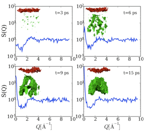
In order to develop a qualitative understanding of these results, comparison has been made to recent MD simulations of the ablation process in Silicon under femtosecond excitation. Silicon and InSb share similar phase diagrams, bonding, and structure so that the MD simulations are expected to be qualitatively similar to that for InSb. Fig. 3 displays snapshots of the near surface region for laser fluences near the ablation threshold, showing the spontaneous development of voids beneath the surface, growing to nanoscale dimensions.
Also shown are the calculated structure factors for each instant in time. One observes the development of a quasi-divergence in the structure factor, as seen in the experimental data, reflecting the development of longer length-scale density fluctuations in the liquid, the first steps in the nucleation of the vapor phase. These measurements thus capture the the nanoscale, ultrafast fluctuation dynamics that underlie superheated liquids, first order phase transitions in materials, and ablation physics, and support theoretical models of the ablation process through MD simulations. Future measurements at the LCLS with coherent beams may be able to capture the shape of the nanoscale void structure, and to investigate the dynamics of nucleation in nanosize materials in which the size of the material is on the order of the size of the critical nucleus.
[1] C.V. Shank, R. Yen, C. Hirlimann, Phys. Rev. Lett., 51, 900 (1983).
[2] A.M. Lindenberg et al., Science, 308, 392 (2005)
[3] K. Sokolowski-Tinten et al., Phys. Rev. Lett. 87, 225701 (2001)
[4] A. Rousse et al., Nature, 410, 65 (2001).
[5] C. Siders et al., Science, 286, 1340 (1999).
A.M. Lindenberg, S. Engemann, K.J. Gaffney et al., "X-ray diffuse scattering measurements of nucleation dynamics at femtosecond resolution", Phys. Rev. Lett. 100, 135502 (2008)


