
X-ray imaging at fine spatial resolution with high compositional and chemical sensitivity is highly desirable in a wide range of scientific and engineering applications. High-profile examples include 1) detection of metal contaminations in Si wafers, which can jeopardize the manufacturing of integrated circuits; 2) electrode dissolution and precipitation in lithium-ion batteries, which can cause performance decay and lead to safety concerns; and 3) metal poisoning in catalytic materials for petroleum refinery, which can significantly affect the energy and economic efficiency of this industry. Generally speaking, the compositional heterogeneities, from the material level to the device level, either purposely engineered or unintentionally formed, have very important implications. Therefore, visualizing the spatial distribution of elements of interest and understanding their behavior are important to both fundamental research and practical applications, very broadly.
Various technical challenges, however, limits X-ray fluorescence imaging’s experimental throughput, dose efficiency, and data quality, hindering its adoption in many of the important applications. A research team led by Dr. Yijin Liu, a previous Lead Scientist at SSRL and currently an Associate Professor in Walker Department of Mechanical Engineering in the Cockrell School of Engineering at UT Austin, developed a novel approach to address the challenges in this field. In contrast to the common endeavor of creating a uniform x-ray illumination, they purposely distort the illumination patterns and use them to encode the specimens’ structural and chemical information, which is later reconstructed using their computer algorithm. This low-cost method utilizes sandpapers as X-rays diffusers, and achieves a spatial resolution down to ~100 nanometer level, which represents a very significant improvement from the previously reports of relevant approaches.
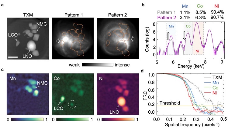
For demonstration of the new method, the authors imaged a composite battery cathode sample composed of a mixture of LiCoO2, LiNiO2, and LiNixMnyCozO2, which are highly relevant to the batteries used in today’s consumer electronics and electrical vehicles (Fig. 1). It is anticipated that this method will open vast scientific and engineering opportunities in various fields as discussed above.
The research team implemented this imaging setup at Beamline 6-2c of SSRL for a proof-of concept demonstration by leveraging and building upon the existing transmission X-ray microscope (TXM) hardware. This work at SLAC is supported by the Department of Energy, Laboratory Directed Research and Development program under contract DE-AC02–76SF00515.
J. Li, S. Chen, D. Ratner, T. Blu, P. Pianetta and Y. Liu, "Nanoscale Chemical Imaging with Structured X-ray Illumination", Proc. Natl. Acad. Sci. USA 120, e2314542120 (2023), doi: 10.1073/pnas.2314542120
