
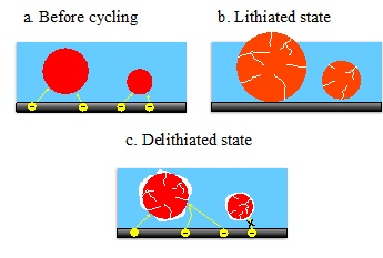
Figure 1. Schematic of the irreversible deformation of the carbon/polymer binder matrix (blue) due to the large Ge (red/orange) volume changes during the first cycle. The deformation results in smaller particles becoming physically disconnected from the conductive matrix, rendering them inactive in subsequent cycles.
In order to successfully replace fossil fuels with cleaner, renewable energy sources, rechargeable battery technology for electric vehicles requires dramatic increases in performance. The lithium-ion battery technology used in portable electronics and electric vehicles has seen only marginal improvements since its commercialization in 1991. New high-capacity electrode materials are required to extend the range of electric vehicles and dramatically reduce their cost. Alloying anodes using silicon or germanium provides excellent high-capacity candidates, however, during cycling these materials are known to undergo dramatic volume changes believed to be responsible for their rapid failure after a few cycles. One possible failure mechanism is the fracturing and eventual pulverization of particles which render fragments electronically isolated.
In a recent study, researchers from SSRL and Stanford University used transmission x-ray microscopy (TXM) during battery operation to observe the volume changes and degradation of micron-sized germanium (Ge) particles during the first two electrochemical cycles. With 2D operando TXM, significant size dependencies in the cycling behavior of Ge particles were observed. During the first cycle, only particles with diameters larger than a few microns showed cracks, and of the ones that show cracking, the smaller particles experienced volume expansion and cracking before their larger counterparts. However, by the second cycle, the smaller particles lost electrical contact, and volume changes were only observed in the largest Ge particles (Figure 1).
Using in situ tomographic TXM at key points along the electrochemical cycle, the first unambiguous evidence of the fracturing of an anode material into completely isolated pieces was established (Figure 2). With the full 3D information, the density and volume changes were calculated. The density changes correlate to a partial transformation into a Li15Ge4-like phase after lithiation and an incomplete delithiation process (Figure 2d).
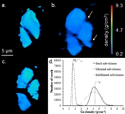
Figure 2. Volume renderings from in situ tomographic imaging of Ge particles (a) before cycling, (b) after the first lithiation cycle, and (c) after the first delithiation cycle. Arrows indicate large cracks formed during the lithiation cycle (b) that fracture the largest particle during delithiation (c). (d) Plot of the Ge density histogram within a 2.2 µm x 2.2 µm subvolume taken from entirely within the top Ge particle. The Ge densities for Ge and Li15Ge4 are displayed as vertical dashed lines. After lithiation, the peak shifts to lower density, corresponding to a ~320% drop in density. The peak falls just shy of the Ge density expected for pure Li15Ge4. After delithiation, the peak center returns nearly to the peak position before cycling, but it has a broad tail towards lower density indicating that many 3D pixels, or voxels, are not entirely delithiated.
This work represents the first direct evidence of the physical and electronic disconnection of active electrode materials during cycling. It also demonstrates the value of linking electrochemical performance with
J. Nelson Weker, N. Liu, S. Misra, J. C. Andrews, Y. Cui and M. F. Toney, "In Situ Nanotomography and Operando Transmission X-ray Microscopy of Micron-sized Ge Particles", Energy Environ. Sci. 7, 2771 (2014), DOI: 10.1039/C4EE01384K
