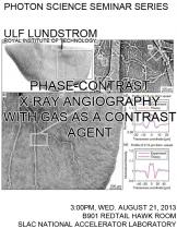
Ulf Lundstrom, Royal Institute of Technology
Conventional X-ray angiography based on iodinated contrast agents is limited to blood vessels no smaller than 50 µm in diameter for radiation doses compatible with living small animals. This is a fundamental limit of absorption contrast set by photon noise, but it can be circumvented through the use of phase-contrast imaging. With propagation-based phase-contrast imaging and carbon dioxide gas as contrast agent we visualize vessels down to 8 µm in diameter at reasonable radiation doses. CO2 is a clinically accepted contrast agent currently used to reduce the absorption in vessels by temporarily displacing the blood. We use a liquid-metal-jet X-ray source to get a small enough X-ray spot with a decent power. The method has been applied to rat kidneys as well as mouse tumors. The low dose requirements indicate a potential for in vivo imaging and longitudinal studies of angiogenesis.


