Renewable energy is critical for the future of humankind. One bottleneck is energy storage because the harvest and consumption of energy are typically separated in time and/or location. Hence, efficient, low-cost, safe and durable batteries are crucial to solve this issue. In order to extend battery cycle life and ensure safety, researchers must understand the evolution of chemical composition and morphology of battery materials during electrochemical cycling.
Lithium-manganese-rich NMC cathode materials, which can be described as Li1+yM1−yO2 (M = Ni, Mn, Co, hence the term NMC), have attracted broad research interest in recent years because of their promising high energy density when cycled at high voltages. However, the battery voltage drops quickly as the electrode is cycled. This critical weakness, known as “voltage fading”, prevents these materials from becoming practical cathode materials.
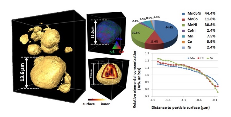
In a recent study, scientists from the University of Science and Technology of China (Feifei Yang and Ziyu Wu), SSRL (Yijin Liu, Joy C. Andrews and Piero Pianetta) and Oak Ridge National Laboratory (Jagjit Nanda, Surendra K. Martha and Gene E. Ice) have investigated the evolution of the battery cathode material Li1.2Mn0.525Ni0.175Co0.1O2 upon cycling. They combined full-field transmission x-ray microscopy (TXM) with x-ray absorption near edge structure (XANES) in order to spatially resolve changes in the chemical phase, oxidation state and morphology of the material. The 3D structure of the cathode particles was retrieved with high spatial resolution and good statistics (>100 particles), revealing elemental/chemical sensitivity in a non-destructive manner (see Fig. 1).

Fig.1 3D morphology of the cathode particles reconstructed from nano-tomography data (left panel). The distribution of the three transition metal ions is rendered and quantified in the middle and right columns.
In this study, spectroscopic data at the Mn K-edge provided a chemical fingerprint, indicating structural changes and/or degradation with high-voltage electrochemical cycling. Single-pixel (~30 nm) TXM-XANES revealed changes in Mn chemistry with cycling, possibly to a spinel conformation and likely including some Mn(II), starting at the particle surface and proceeding inwards.
Tomographic reconstruction of cathode particles cycled once and 200 times also showed significant variations in particle morphology, with the majority of particles adopting non-spherical shapes when cycled.
Further analysis via multiple-energy tomography revealed that transition metals were approximately 80 percent homogeneously distributed in pristine electrodes but began segregating after just one cycle – an effect that became larger after 200 cycles. The researchers then analyzed the bulk 3D elemental transition metal distribution and its variation from particle center to surface, taking into account changes in internal geometry that produced surface-like structures within the particles. This depth dependency of the elemental associations showed depletion of transition metals at outer and inner cathode surfaces after just one cycle, driven by electrochemical reactions at the surface, suggesting that inner macro pores are likely connected to the particle surface through nanoscale pores or cracks.
Besides the important scientific findings in this paper, the authors also demonstrated the powerful capability of their method. They expect TXM-XANES to further develop and become increasingly influential as more and more scientific studies will make use of this novel method.
F. Yang, Y. Liu, S. K. Martha, Z. Wu, J. C. Andrews, G. E. Ice, P. Pianetta and J. Nanda, "Nanoscale Morphological and Chemical Changes of High Voltage Lithium–Manganese Rich NMC Composite Cathodes with Cycling", Nano Lett. (2014), DOI: 10.1021/nl502090z


