
Contents of this Issue:
1. Science Highlight —
Novel Ferroelectric Nanostructures for Nanoelectronic Devices
(contacts: L.E. Fuentes-Cobas, Advanced Materials Research Center)
 | |
| Experimental 2-D diffraction pattern. |
To learn more about this research see the full scientific highlight at:
http://www-ssrl.slac.stanford.edu/research/highlights_archive/fuentes.html
2. Science Highlight —
The Structure of a Reaction Intermediate in Enzymatic
Halogenation
(contact: P. Riggs-Gelasco, College of Charleston)
Halogenated natural products play important roles as antibiotics, antifungals,
and antitumor agents. The process of halogenation involves the replacement of a
hydrogen with a halide (such as chloride or bromide), and is a challenging task
for a synthetic chemist. However, the iron-containing enzymes in the
haloperoxidase and halogenase families readily catalyze these reactions. It is
thought that when this reaction occurs, the iron in the enzyme is at a
high-valent Fe(IV) state, and that this species is responsible for removing a
hydrogen atom (called an abstraction) from the substrate, creating a substrate
radical, and that a halogen radical is subsequently transferred to the
substrate to complete the halogenation reaction. Recently, Pamela Riggs-Gelasco
and co-workers used x-ray absorption spectroscopy at SSRL's Beam Line 7-3 to
obtain unique structural insights into this enzymatic intermediate in the
halogenase CytC3. The extended x-ray absorption fine structure (EXAFS) data
showed the presence of a short Fe-O bond and a Fe-Br interaction, clearly
identifying a Br-Fe(IV)=O2- unit and confirming a key component in
the enzymatic mechanism.
To learn more about this research see the full scientific highlight at:
As you know, since January there have been discussions about how the reduced
budget will impact SSRL activities for this fiscal year. SSRL Lab Management
has made the decision to continue the current user run as originally projected
- ending at 6 am on Monday, August 11. However, there will be a 2-1/2 week
shutdown June 30-July 15, 2008. During this period, we have scheduled
Maintenance/Accelerator Physics (MA/AP) so that equipment related to top off
injection can be installed. Other activities which would normally disrupt user
operations may also be scheduled during this shutdown, such as seismic retrofit
construction needed in Building 120.
Modern synchrotron radiation based x-ray absorption spectroscopy (SR-XAS)
techniques offer the ability to probe local molecular scale physical and
electronic structures that govern key properties of technological and
environmental materials and molecular complexes. The high collimation,
intensity, and tunability of SR allow the investigation of a wide range of
materials, including thin films and interfaces, nanoparticles, amorphous
materials, solutions, hydrated and disordered minerals, soils, and dissolved
species.
7.
2008 SSRL/LCLS Users' Meeting, October 15-18, 2008
Mark your calendar for the second joint SSRL and LCLS Users' Meeting and
Workshops on October 15-18, 2008. Several parallel sessions for SSRL and LCLS
science as well as joint sessions to discuss topics of general interest and to
feature science highlights from the last year are being planned. SSRL specific
workshops on October 15, will include: 1) Advanced Topics in EXAFS Analysis and
Applications, 2) Macromolecular Crystallography, and 3) a joint ALS/SSRL
workshop on Introduction to Synchrotron Radiation Experiments. LCLS workshops
on October 17-18 will include: 1) Coherent X-ray Scattering and Imaging; 2)
AMO/Strong Field Control of X-ray Processes, and 3) Soft X-ray Instrumentation
for LCLS.
__________________________________________________________________________
SSRL Headlines is published electronically monthly to inform SSRL users,
sponsors and other interested people about happenings at SSRL. SSRL is a
national synchrotron user facility operated by Stanford University for the
U.S. Department of Energy Office of Basic Energy
Sciences. Additional support for
the structural biology program is provided by
the DOE
Office of Biological and Environmental Research, the NIH
National Center for Research Resources and the NIH Institute for General Medical
Sciences. Additional information about
SSRL and its operation and schedules is available from the SSRL WWW
site.
__________________________________________________________________________
To leave the SSRL-HEADLINES distribution, send email as shown below:
To: LISTSERV@SSRL.SLAC.STANFORD.EDU
Subject: (blank, or anything you like)
The message body should read
SIGNOFF SSRL-HEADLINES
That's all it takes. (If we have an old email address for you that is
forwarded to your current address, the system may not recognize who
should be unsubscribed. In that case please write to
ssrl-headlines-request@ssrl.slac.stanford.edu and we'll try to figure out
who you are so that you can be unsubscribed.)
If a colleague would like to subscribe to the list, he or she should send
To: LISTSERV@SSRL.SLAC.STANFORD.EDU and use the message body
SUBSCRIBE SSRL-HEADLINES
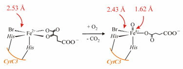
The reaction catalyzed by the halogenase CytC3.
http://www-ssrl.slac.stanford.edu/research/highlights_archive/halogenasecytc3.html
3.
Budget Impact on SSRL User Operations
(contact: J. Stöhr, stohr@slac.stanford.edu)
Scheduling this shutdown in July will likely result in reduced power and labor
costs, but more importantly, utilizing the down time to install and test top
off equipment provides the opportunity of better understanding how the top off
mode will impact beam line instrumentation, optics and user experiments before
the next run begins in the fall. This will provide valuable information to
proceed with the reviews and approvals needed for more frequent top off mode on
all beam lines in the future. Results from these preliminary tests could then
be shared with the user community at the next Users' Meeting in October.
Pending approval from DOE and SLAC Radiation Physics (RP), top off mode
operation may be continued during the remainder of the run (July 16-August 11)
on selected beam lines on the existing fill schedule of injection every 8 hours
(but with shutters open on selected beam lines). RP may designate an 'exclusion
area' around the selected beam lines and those areas may need to be cleared
during the daily top off fills at 6 am, 2 pm, and 10 pm (fills usually take ~5
minutes). Little impact to user operations is anticipated as a result of these
tests.
SPEAR Operating
Schedule
4.
Very Small Spot Size Achieved at the New BL13-2
(contact: H. Ogasawara, hirohito@slac.stanford.edu)
SSRL staff have been making progress on the soft x-ray emission spectroscopy
capability at the new BL13-2 (first light at BL13 - the newest of SSRL's beam
lines - and the description of its end stations - was reported in last month's
SSRL Headlines). Soft x-ray emission spectroscopy is receiving growing
attention for its unique ability to provide an atom-specific picture of the
electronic structure in matter. Soft x-ray emission spectroscopy allows studies
of many different samples: solids, surfaces, gases and liquids. The method is
well suited to study gases and liquids and their solid interfaces since it is
an x-ray-in/x-ray-out technique that does not require high vacuum. A typical
soft x-ray emission spectrometer consists of an entrance slit, a spherical
grating, and a 2D-detector. The entrance slit is the source point: a wide
entrance slit gives high detection efficiency with low energy resolution,
whereas a narrow entrance slit gives high energy resolution but low detection
efficiency. SSRL staff is developing a high throughput x-ray emission
spectrometer without an entrance slit and plan to use an extremely tightly
focused beam at the sample on BL13-2. In this design, the detection efficiency
will only depend on the acceptance angle of the grating and thereby the energy
resolution will be determined by the size of the focal spot at the sample. To
obtain typical resolving power (D E/E) of about
1000, the beam needs to be focused down to 10-20 microns along the dispersive
direction (vertical). At the new BL13-2, a pair of Kirkpatick-Baez refocusing
mirrors image the source horizontally and the exit slit vertically onto the
sample. On March 22, SSRL staff achieved a key milestone during the current
commissioning by measuring a beam size smaller than 5 microns in the vertical
and 60 microns in the horizontal dimension at the sample. When fully
commissioned, BL13 and its three stations will be opened as a general user beam
line.
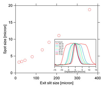
Vertical spot size
measurement at BL13-2 as a function of exit slit size. [larger version]
5.
SSRL School on Synchrotron X-ray Absorption Spectroscopy Techniques in
Environmental and Materials Sciences: Theory and Application
(organizers: S. Webb, J. Bargar, M. Toney and A. Mehta)
Good planning and a working knowledge of beam lines, in addition to techniques,
are keys to conducting successful SR-XAS measurements. This third annual school
on SR techniques will provide a practical users' guide to planning and
conducting XAS/EXAFS measurements at SSRL beam lines. The school will cover
important basics such as basic beam line setup and optimization for
fluorescence and transmission spectroscopy, as well as advanced techniques
including polarized single-crystal EXAFS, grazing incidence XAS, and microprobe
XAS.
We will cover topics that are not commonly addressed in text books or class
lectures, but are obtained only through on-the-experiment training. Tips on
sample preparation, data collection, processing and analysis will be included.
The first day of the school (May 20) will be a lecture day, followed by
practical days with a full hands-on day at SSRL beam lines (May 21) and a third
day of data analysis and discussion (May 22). Registration is limited, so
please register early. Cost will be $75.
http://www-ssrl.slac.stanford.edu/conferences/workshops/srxas2008/index.php
6.
Ultrafast X-ray Summer School, June 17-20, 2008
(SLAC Today article by M. Cunningham)
The Linac Coherent Light Source (LCLS) will enable scientists to explore new
chemical structures and processes. But before scientists grab the reins in this
novel venture they'll have to go to school-Ultrafast X-ray Summer School, that
is.
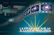
The Ultrafast X-ray Summer School, which is slated to run June 17-20, will
hinge upon participant-oriented discussion and interaction focused on the
state-of-the-art science that the LCLS will set in motion.
"There is a need to educate both senior scientists and younger scientists to
generate a new class of researcher that will utilize this unique opportunity,"
Chairman Kelly Gaffney said. "And, for at least the next couple of years, this
is the only place in the world it will be possible."
The LCLS has properties traditionally utilized in the laser community, but
because it generates x-rays, it presents new opportunities. Techniques used in
laser science and synchrotron x-ray science typically don't overlap-but the
success of the LCLS will depend on the merging of these fields and the skill
sets of each.
LCLS x-rays are expected to provide instantaneous images of atomic and
molecular structures, which require a camera with sub-nanometer spatial
resolution and a shutter speed of less than a trillionth of a second. This will
generate unique opportunities for capturing single bimolecular structures and
collecting real-time movies of chemical, physical and biological
transformations.
Interested applicants can find registration and program information at the
workshop website. Rooms at the Stanford Guest House are expected to fill
quickly, so attendees are encouraged to make their reservations soon.
http://www-conf.slac.stanford.edu/uxss/2008/
Contact the meeting organizers if you have suggestions for the annual meeting:
Wayne Lukens (LBNL), Amyeric Robert (LCLS/SLAC), Riti Sarangi (SSRL/SLAC), and
Linda Young (ANL). Start to think about topics which you may want to share
through contributed talks or poster presentations; and consider nominating your
colleagues for awards which will be presented at the annual Users' Meeting. The
call for abstracts and award nominations will follow shortly. To review the
2007 meeting and workshop report, see
http://www-ssrl.slac.stanford.edu/pubs/2007_ssrl_lcls_meeting_report.doc
http://www-conf.slac.stanford.edu/ssrl-lcls/2007/2008.htm
8.
Big Science Gets Small: New BL6-2 Microscope
(SLAC Today article by B. Plummer)
The size of big science at the Stanford Synchrotron Radiation Laboratory (SSRL)
just got a little smaller, thanks to a new x-ray microscope installed last fall
at Beam Line 6-2. As one of only a handful of such instruments in operation
around the world, the new microscope offers SSRL researchers and users a new
set of eyes for peering into the three-dimensional chemistry and structure of a
range of materials from meteorites to mouse bones.
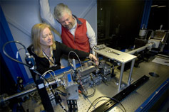
Joy Andrews and Sean Brennan with the new nano-imaging x-ray microscope
at BL6-2.
Spearheaded by Piero Pianetta and built by x-ray equipment supplier Xradia, the
otherwise commercially available microscope takes advantage of the SPEAR3
synchrotron's incredible brightness to quickly capture three-dimensional images
of structures as small as 40 nanometers, or about the size of the smallest
virus.
"It's just like a typical microscope, but instead of looking with light, which
has a relatively large wavelength, we're looking with x-rays, which have a very
small wavelength," said Joy Andrews, SSRL staff scientist. "That way we can
look at much smaller things."
In one recent study, Andrews and colleagues investigated the effects of
weightlessness on the bones of mice to gather clues about the long-term effects
of spaceflight on humans. Because of the high brightness of the SPEAR3
synchrotron beam, scans of bone tissue can be completed in a fraction of the
time needed for similar studies using other x-ray sources.
The microscope works by passing a thin beam of x-rays through the sample, then
through a special lens called a "zone plate" that enlarges the image onto a
special screen. This special "phosphor" screen uses the same basic technology
as a traditional television screen to convert the invisible x-ray image into a
visible image which is then picked up by a specially designed digital camera.
The new microscope has the added advantage of using tunable x-rays that can be
changed to reveal the chemistry of a sample, giving researchers the power to
scrutinize materials to a level impossible without a synchrotron x-ray source.
To obtain a final, three-dimensional image, the sample is scanned dozens of
times, rotating a fraction of a degree with each shot. The images are combined
to construct a movie that reveals the nanoscale structural features of the
sample.
SSRL Welcome
Page | Research
Highlights | Beam Lines | Accel
Physics
User
Admin | News & Events |
Safety Office