
Contents of this Issue:
1. Science Highlight —
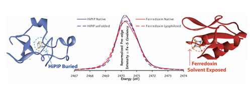
Tuning the Properties of Iron-Sulfur Clusters in Proteins
(contacts: B. Hedman, hedman@ssrl.slac.stanford; K.O. Hodgson,
hodgson@slac.stanford.edu; E.I. Solomon, edward.solomon@stanford.edu)
Proteins containing iron-sulfur clusters are ubiquitous in nature and catalyze
one-electron transfer processes. These proteins have evolved into two classes
that have large differences in their electrochemical potentials: high potential
iron-sulfur proteins (HiPIPs) and bacterial ferredoxins (Fds). The role of the
surrounding protein environment in tuning these redox potentials has been a
persistent puzzle in the understanding of biological electron transfer.
Although high potential iron-sulfur proteins and ferredoxins have the same
iron-sulfur structural motif, there are large differences in their
electrochemical potentials. HiPIPs react oxidatively at physiological
potentials, while Fds are reduced. Sulfur K-edge x-ray absorption spectroscopy
(XAS; measured at SSRL Beam Line 6-2) has been used to uncover the substantial
influence of hydration on this variation in reactivity, in a collaborative
effort led by Stanford Chemistry and Photon Science researchers. The study
showed that the Fe-S covalency (a measure of the electronic overlap of the
sulfur and iron orbitals forming the bonds within the clusters) is much lower
in natively hydrated Fd active sites than in HiPIPs, but increases upon water
removal; similarly, HiPIP covalency decreases when reversibly unfolded,
exposing an otherwise hydrophobically shielded active site to water. These
results demonstrate that the Fe-S covalency determined from the sulfur-K XAS
data is a direct experimental marker of the local electrostatics due to
H-bonding. Studies on related model compounds and accompanying density
functional theory calculations support a correlation between Fe-S covalency and
ease of oxidation, which suggests that differential hydration accounts for most
of the difference between Fd and HiPIP reduction potentials. This raises the
intriguing possibility that oxidation/reduction potentials can be regulated by
protein/protein and protein/DNA interactions that effect cluster hydration.
To learn more about this research see the full scientific highlight at:
In preparation for a key program review of SSRL by the DOE Office of Basic
Energy Sciences and for our Annual NIH, NCRR/Biomedical Technology Program
(BTP) Progress Report, SSRL needs your support. Please provide us, by December
5, with a list of publications, major awards and invited lectures for work
based at least in part on use of SSRL resources for years 2005-2007. Please
send your list of publications by email to lisa@slac.stanford.edu or by using
the web form at:
http://smb.slac.stanford.edu/forms/reporting/form_publication.shtml.
Please also flag those that you view as being "high profile, high impact" with a
sentence explaining the impact.
4.
JCSG Celebrates Its 500th Structure
SSRL has contributed to the development of robotic hardware to process and
evaluate the crystal samples, enabling fast sample turn-around at the beam
lines, and has generated novel software programs for high through-put structure
determination. "Because the beam line is fully automated, we can control
almost every step in our experiment from any computer," said Jessica Chiu, who
manages crystal screening and data collection activities at SDC. "We can be at
home, or in another country, and still screen crystals around the clock. This
allows us to attain maximal throughput at the beam lines with minimal human
intervention."
The determination of 500 structures is a tremendous accomplishment, and a
testament to the talented scientific team and to the success of their
automated, highly efficient operation. "We've doubled the rate at which we
solve new structures since 2005," Deacon reported, "and we're still getting
faster. But we won't compromise quality. The best way to speed things up is to
get the best quality data you possibly can."
More information on all of the structures solved and deposited by the JCSG can
be found at the JCSG Structure Gallery.
JCSG is funded by the NIH National Institute of General Medical Sciences,
Protein Structure Initiative U54GM074898. http://www.jcsg.org/
There are new procedures related to international shipments, so please contact
us in advance if you are planning to ship samples, equipment or other
scientific items from SSRL to a location outside of the US following your beam
time at SSRL (for example, to return dewars to a collaborator or your home
institution).
__________________________________________________________________________
SSRL Headlines is published electronically monthly to inform SSRL users,
sponsors and other interested people about happenings at SSRL. SSRL is a
national synchrotron user facility operated by Stanford University for the
U.S. Department of Energy Office of Basic Energy
Sciences. Additional support for
the structural biology program is provided by
the DOE
Office of Biological and Environmental Research, the NIH
National Center for Research Resources and the NIH Institute for General Medical
Sciences. Additional information about
SSRL and its operation and schedules is available from the SSRL WWW
site.
__________________________________________________________________________
To leave the SSRL-HEADLINES distribution, send email as shown below:
To: LISTSERV@SSRL.SLAC.STANFORD.EDU
Subject: (blank, or anything you like)
The message body should read
SIGNOFF SSRL-HEADLINES
That's all it takes. (If we have an old email address for you that is
forwarded to your current address, the system may not recognize who
should be unsubscribed. In that case please write to
ssrl-headlines-request@ssrl.slac.stanford.edu and we'll try to figure out
who you are so that you can be unsubscribed.)
If a colleague would like to subscribe to the list, he or she should send
To: LISTSERV@SSRL.SLAC.STANFORD.EDU and use the message body
SUBSCRIBE SSRL-HEADLINES
Schematic representation of the common active-site iron-sulfur cluster
structural motif.
http://www-ssrl.slac.stanford.edu/research/highlights_archive/iron-sulfur_clusters.html
2. Science Highlight —
A Step Toward Understanding High-Temperature Superconductors
(contacts: D.H. Lu, dhlu@slac.stanford.edu; Z.-X. Shen, zxshen@stanford.edu)
Scientists can make high-temperature superconductors, but they don't have a
good theory for how they work. Understanding high-temperature superconductors
will have significant impact on the modern condensed matter theory, and may
someday allow scientists to design room-temperature superconductors. SLAC
Photon Science and Stanford Professor Z.-X. Shen and colleagues, working at
SSRL's Beam Line 5-4, recently made observations that will help shape the
theory. Their results are published in the Nov. 1 issue of Nature.
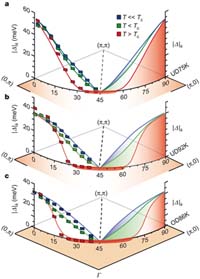
a,
Underdoped sample with Tc = 75 K. b, Underdoped sample
with Tc = 92 K. c, Overdoped sample with
Tc = 86 K. At 10 K above Tc there exists a
gapless Fermi arc region near the node.
[see larger image and more detailed caption at Nature].
Certain materials become superconductors-that is, losing all resistance to
electric current-when they become colder than their transition temperature. So
far, the warmest transition temperature recorded is minus 164 degrees
Fahrenheit. Below the transition temperature, the electrons pair up. This
pairing reduces the energy of the electrons. The strength of pairing is
characterized as a superconducting gap in the single particle excitation
spectrum, which is found below the transition temperature in both conventional
and high-temperature superconductors.
In high-temperature superconductors, scientists also observe a gap above the
transition temperature, called the pseudogap. So far it has been unclear
whether the two gaps are unrelated, or if the pseudogap is a precursor to the
superconducting gap. Using angle-resolved photoemission spectroscopy to
measure the energy gap at different temperatures and momenta, Shen and
colleagues found that the pseudogap and the superconducting gap coexist and
exhibit different temperature dependence. Therefore, the two gaps seem to have
different origins. This should provide an important step toward unveiling the
mystery of the pseudogap phenomena.
http://www-ssrl.slac.stanford.edu/research/highlights_archive/high-tc_07.html
3.
Call for User Publications, Awards, Invited Lectures
(contacts: L. Dunn, lisa@slac.stanford.edu)
Lists of publications reported to SSRL to date and sample acknowledgment
statements can be found at: http://www-ssrl.slac.stanford.edu/pubs/
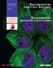
Biochemistry and Cell Biology December cover image
(85: 537-542 (2007)).
Inset image: Synchrotron X-ray fluorescence mapping reveals the asymetric
distribution of iron in an intact Xenopus laevis oocyte.
Image: Popescu et. al, University of Saskatchewan Background image: Human
breast cancer cells MCF-7 immunostained for microtubules (alpha-tubulin) in
green and transcription factor Sp1 in red, with nuclei in blue. Image provided
by Shihua He, University of Manitoba.
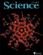
Science October 19 cover
image (318, 430 (2007)).
Structure of a gold nanoparticle in which the central atoms are packed in a
decahedron, surrounded by additional layers of gold atoms in unanticipated
geometries. Gold atoms, gold; sulfur atoms, blue; carbon atoms, white; oxygen
atoms, red; the superimposed red mesh depicts the electron-density distribution
determined by x-ray crystallography. Image: Pablo D. Jadzinsky
and Guillermo Calero.
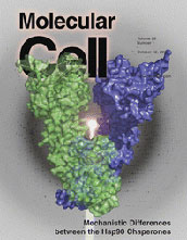
Molecular Cell October 12 cover image
(28, 41 (2007)).
The high-resolution structure of the mammalian endoplasmic reticulum Hsp90
chaperone GRP94 with bound ATP. The extent to which a common mechanism applies
to all Hsp90 chaperones has been controversial. In cytosolic Hsp90s, ATP
binding results in N-terminal dimerization that is critical for ATP hydrolysis
and subsequent chaperone function. Dollins et al. show that
nucleotide-bound GRP94 adopts a conformation that precludes N-terminal
dimerization yet demonstrate GRP94-catalyzed ATPase activity using kinetic
analyses, suggesting that nucleotide binding is not the major driving force for
catalytically productive conformational changes.
—SLAC
Today article by Elizabeth Buchen
On October 29, the Joint Center for Structural Genomics (JCSG)-in which SLAC
plays an integral role-celebrated a major milestone as it deposited its 500th
unique protein structure into the Protein Data Bank (PDB). A protein's 3D
structure dictates its function-including how it interacts with other proteins,
how it catalyzes chemical reactions and how it might be inhibited or activated
by drugs. By determining structures representative of large protein families
from a wide range of genomes, the JCSG seeks to illuminate key aspects of
biology, chemistry and medicine that span from neurodegenerative disease to
human evolution.
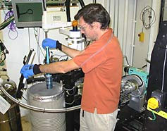
Ashley Deacon loads samples of crystallized proteins into an automated sample
mounting system.
The process of depositing a protein structure into the PDB begins with the
Bioinformatics Core (based at the University of California San Diego and the
Burnham Institute for Medical Research) selecting the targets to be
investigated and the Crystallomics Core (based at The Scripps Research
Institute and the Genomics Institute of the Novartis Research Foundation)
producing soluble proteins that are then crystallized. Researchers at SLAC's
Stanford Synchrotron Radiation Laboratory (SSRL) then use x-ray diffraction to
map out the atomic structure of each crystallized protein. This Structure
Determination Core (SDC) group determines the 3D structures and deposits the
structural coordinates and diffraction data into the PDB, where they are freely
available to the scientific community.
"Together, the Institutes in our consortium have established a very effective
pipeline," said SLAC's Ashley Deacon, who leads the SDC. "We have developed
new technologies and automated the various steps of the process to scale up not
only production, but also to increase the efficiency, lower the cost and
increase the quality of the structures determined."
5.
Planning Any International Shipments?
(contacts:
C. Knotts, knotts@slac.stanford.edu; L. Dunn, lisa@slac.stanford.edu)
7.
User Administration Update
(contact:
C. Knotts, knotts@slac.stanford.edu)
Macromolecular Crystallography Proposals due December 1: If you are interested
in submitting a proposal for the December 1, 2007
deadline for time on
macromolecular crystallography beam lines, see:

http://smb.slac.stanford.edu/public/forms/becominguser/
X-ray/VUV Beam Time Requests due December 7: Please submit your requests for
X-ray/VUV beam time for the February-May 2008 scheduling period.
http://www-ssrl.slac.stanford.edu/users/user_admin/xray_btrf.html
http://www-ssrl.slac.stanford.edu/users/user_admin/vuv_btrf.html
Start your holiday shopping on your next visit to SSRL: When you check in for
beam time, check out our Texas Orange t-shirts commemorating the first
joint SSRL and LCLS users' conference. On sale now - just $10 - while supplies
last. See Michelle Steger Bldg. 120, Rm. 219.
SSRL Welcome
Page | Research
Highlights | Beam Lines | Accel
Physics
User
Admin | News & Events |
Safety Office