|
|
Beamline 11-2 Operators Manual
Table of Contents:
- Beamline Source
- Beamline Optics
- In Hutch Equipment
1. Beamline Source
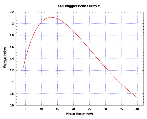
Beamline 11-2’s source is a 26 pole, 2 Tesla hybrid magnet wiggler with a 175 mm period which produces these total photon and total power spectra:
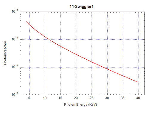
Beamline 11-2 has a horizontal fan of 1.50mrad with an center offset 1.00mrad from the wiggler centerline towards SSRL. Within this fan the x-ray flux is approximately flat from 5KeV through 25KeV. Beamline 11-2 accepts approximately 20% of the photons generated in the wiggler.
2. Beamline Optics
- Description:
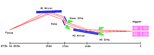
There are three primary optics in the 11-2 configuration; M0 collimating mirror, Monochrometer, and M1 focusing mirror. Each serves a specific task in configuring the beam delivered to the hutch.
The M0 collimating mirror (M0) is a single, 1 meter piece of flat polished (to 0.5nm) Silicon. It is coated with about 40nm of Rhodium, a high density metal, to increase it’s reflectivity at higher incidence angles. This mirror is upward reflecting and has a bender mounting to effect collimation on the incoming beam. In addition, its incidence angle and vertical position are motor driven.
The 11-2 monochrometer is the prototype of its kind at SSRL. It has a liquid nitrogen recirculation system which maintain the mono crystals at about 80K. The advantage to this system is that silicon, fortuitously, goes through a minimum in it’s expansion coefficient curve at these temperatures i.e., the crystal is less likely to distort under heat loads. The beamline 11-2 monochrometer has the highest energy resolution of any similar mono at SSRL. It is upward reflecting with a 3 inch primary crystal and a 3.75 inch secondary separated by 6mm and capable of reflecting a full 2mm high beam at angles from 5 to 45 degrees.
The M1 mirror is also a 1 meter piece of polished (to 0.5nm) Silicon but it has a sagittal radius ground into the top surface. This allows the mirror, when bent, to approximate the outer surface of a toroid, the required shape for focusing a point source in this geometry. It is a downward reflecting mirror and, like M0, also coated with 40nm of rhodium. In addition to the mirror bender, there are motorized pitch, vertical translation, and mirror yaw controls.
The mono and the M1 mirror are mounted in sequence on a single large motorized blue girder in the 11-0 hutch. In this fashion, a change to the pitch on M0 can be accommodated by pitching both devices at once.
In addition to the primary optics, there are a number of other devices in the beam. Vertical slit aperture the beam in front of the M0 mirror. These are required to prevent the mirror from taking direct beam anywhere other than it’s top surface. Vertical and horizontal slit jaws are located in front of the mono. The vertical jaws aren’t typically used except during beam checkout but the horizontal jaws are the primary method of reducing flux on a experimental sample. Just downstream of the mono is a moveable diagnostic foil which can be useful during checkout tests but is never used during experiments. A general diagram of the beamline optics is shown below.
- Optics Function:
Mirror Focus along the beam follows the form:
Rtan = (2*Fi*Fo)/(a*( Fi+Fo))
Looking at the dimensions of beamline 11, the wiggler to M0 distance is 14 meters but ideally we would like an infinite focus coming out of the mirror. This gives a tangential radius of:
Rtan » 2/a
Where a is the pitch angle in radians. For a typical cutoff energy of about 14KeV the a value on 11-2 is about 4.7mrad so the bend radius required is around 425m. The same tangential equation describes the vertical focusing of the parallel light coming off the monochrometer by the M1 mirror.
The M1 mirror with it’s ground sagittal radius is designed for optimal focus at a point approximately 10 meters from the mirror. This places the best focus position in the center of the rear hutch table. For other sample positions, the mirror needs to be over-bent to bring the vertical focus closer to the mirror. This leaves the sagittal radius, which is fixed, as inadequate for the horizontal hocus. The result is that the beam image appears as a frown or slightly butterfly shaped. The resulting image dimensions achieved range from a best case of 0.8mm W x 0.2mm H to the highest pitch setting at the front table of about 2.2mm W x 0.4mm H.
- Configuring the Optics:
The first choice to be made in deciding how to set up 11-2 for an experiment is determined by the experiment itself. What is the concentration of the species to be measured? What is the susceptibility of the samples to photo reduction or x-ray damage in the beam? How uniform are the samples, both in thickness and concentration? Answers to these questions will determine what configuration to put the optics in.
There are three primary ways to set up the Beamline 11-2 optics; uncollimated, unfocused, and focused. Uncollimated refers to a setup like Beamline 4-1, direct beam from the wiggler to the mono. Possibly filters and slits will be used to aperture the beam. Unfocused refers to the M0 mirror being in to set an energy cutoff while the M1 mirror is left out, leaving the hutch beam approximately parallel. The focused condition refers to both mirrors in.
What are the advantages and disadvantages to each configuration?
First, the uncollimated condition. This setup provides the lowest flux density available. Beamline 11-2 has horizontal and vertical fan widths of 1.5mrad and 0.15mrad so the beam size can become very large at the back hutch table, 29.5m from the source, 44mm wide by 4.4mm high. In this condition about 75 to 80 percent of the source flux is thrown away, a potential problem for samples of low concentration. This arrangement also has the lowest energy resolution. The LN2monochrometer is Darwin width limited for most of it’s energy range. Hence, the vertical fan is larger than the crystal resolution and the energy spread in the uncollimated arrangement is large compared to the unfocused and focused configurations. Another purely practical disadvantage is that this setup provides direct beam on the mono, increasing LN2 consumption. On the plus side, this arrangement is very stable and familiar to most users. Detuning is required to remove higher order harmonic content from the hutch beam. This is the only arrangement usable for energies above 22KeV due to the rhodium coating on the mirrors and the beamline optics design parameters for M0 mirror pitch. It may be the best arrangement for samples which are known to be non-uniform in thickness or homogeneity.
The collimated arrangement provides improved energy resolution and a parallel beam in the hutch. The flux density is higher than the uncollimated configuration but lower than fully focused. Detuning the monochrometer is not required. Beam size in the hutch is about 21mm wide by 2.2mm high. This configuration would be best for samples sensitive to high flux densities but which also require high energy resolution. The collimated setup is not currently used due to pathologies in the M0 mirror which leave it’s profile (hence beam position) susceptible to variations in cooling water temperature and SPEAR current.
The focused configuration provides equal energy resolution to the collimated configuration and the highest flux density available at SSRL. Total flux is about the same as the collimated arrangement but the focus size ranges from 200um high by 800um wide at the back hutch table at the optimal design configuration up to about 600um high by 3000um wide at the highest pitch (7mrad) configuration on the front hutch table. The monochrometer is kept fully tuned. In this arrangement the total flux on the sample is controlled by the width of the horizontal slits in front of the mono. This configuration is best for low concentration samples, samples which are granular (assuming accurate positioning on the species desired), small natural samples and any focusing optic arrangement.
Consult with the SSRL scientific and engineering staff regarding specifics of your experiment to select the best optics arrangement.
3. In Hutch Equipment
- Description:
Beamline 11-2 has many possible hutch configurations available for experimental setup. The first and most obvious difference between 11-2 and other SSRL beamlines is the dual hutch table setup. This arrangement accomplishes two purposes. First, it allows for one table to be tented for actinide or hazardous material analysis. Second, as the M0 mirror cutoff angle increases, the optimum focus for 11-2 moves further up the beamline. This feature leads to the front table as the optimum position for low energy focused and uncollimated experimental conditions.
The two hutch tables are identical in design and construction. They have three separate motors on the which allows for the tables to move up and down, have –2 to +10 mrad pitch capability and can translate ±25mm in the horizontal perpendicular to the x-ray beam. This allows a great deal of flexibility in positioning experimental setups.
The standard rail at 11-2 is designed around a standard x-ray beam height of 11 inches above the table surface. The table is aligned in the vertical and pitch to parallel the beam at this height above the surface. The rail is similar in concept to other in hutch rails at SSRL with some minor differences. It is based upon an Oriel dove tail optical rail which is very stiff. The slits, ion chambers and other in-beam devices are mounted on slides and can be positioned anywhere on the rail. The sample positioner is mounted on a bridge over the rail and allows for vertical and horizontal positioning.
Standard setup on the rail includes a motorized set of four jaw slits, three standard (15cm) ion chambers, a sample box and both Lytle and 30 element germanium detectors. A number of options are available for rail installation: Long (30cm) ion chambers, He flight tubes, a beam imaging camera, additional set of four jaw slits, LHe cryostats, M1 mirror pitch feedback and a filter box are on hand and more equipment will be developed as needs arise.
The rear hutch table is fully equipped for running radioactive or hazardous samples. It is enclosed in a permanent containment tent and has installed HEPA filters and blowers to maintain filtered air flow and monitoring.
Outside the hutch, to the left of the door is the hutch gas control panel which supplies the gas needs for all devices in the hutch. Since this is a focused beamline, electron-ion recombination in the ion chambers is a potential problem. The solution to this is mixing a heavier gas such as argon with helium as a carrier to reduce the beam absorbed in the ion chamber and thus the space charge density and chances of significant recombination. The flowmeters labeled I0, I1, and I2 allow stable and fairly precise mixing of the available gases. In addition, there are controls for Lytle detector and mirror pitch feedback charging and for the helium purge of any installed flight path or chamber. An optional channel is available for any special application.
A primary feature of beamline 11-2 is the 30 element germanium solid state detector. Similar to other Ge detectors, this system has the additional feature of an added articulated element/preamplifier assembly. The detector mount is constructed so that the detector elements can be oriented horizontally or vertically, depending upon the needs of the experiment.
The experimenters station at 11-2 is a standards SSRL setup with a second ion chamber repeater on the partition to the right of the VMS monitor. The left hand of the electronics cabinets contains the beamline personnel protection system as well as a CCTV monitor and the cryostat controls. The right hand enclosure holds al the interface electronics and control systems. The VtoF converters, hex scalers, and ion chamber repeaters are also located here.
In addition, the following data collection equipment which is available to beamline 11-2 experimenters:
- Thermoelectrically cooled sample holder which can maintain a sample at 0 °C in a standard Lytle box sample holder.
- GSK 2001 Lauie secondary WDS system. Consult with SSRL before regarding applications.
- High precision grazing incidence XAFS spectrometer.
- Video imaging cameras.
- Electron yield detector
- Microfocusing capillary and stage for 5µm spot size analysis.
- Alignment and Use:
Prior to arriving at beamline 11-2 for an experimental run, contact the MES staff at SSRL to confer about the best setup for your experiment. A discussion about the type of samples, activity levels, stability, concentration and any special sample or experiment needs will help greatly in selecting the proper configuration for the experiment.
SSRL engineers will setup the beamline optics for the experimenters needs. The sample rail, ion chamber gases and fluorescence detectors will be set up and configured along with any ancillary equipment such as He flight tubes or LHe cryostats.
Once the beam optics have been configured, the first step in aligning the tables is to center beam in the ion chambers. Set the rail slits to about 1 x 1 cm, back out the sample holder and scan the table vertically. Center the beam in I0. Check the position of the beam in I1 and I2 to see if it coincides with the beam position of I0. If the beam profiles are not as wide as the I0 profile, the table is tilted relative to the beam. One edge of the scans will align, the edge where I0 occludes the I1 and I2 detectors. If this edge is the lower or left side of the scan, the table is tilted too much. If the right side coincides in all detectors, increase the table tilt. Once the table tilt is set, do a horizontal table scan to center in I0 and reset the slits to their desired size.
Currently the Ge30 detector motion is not linked to the sample horizontal positioner. Consequently, the sample positioner should be centered in the beam and left there for measurements. This is accomplished by doing a sample horizontal scan with the rail slits set to about 15mm. A sample vertical scan should be done first to assure beam is through to I1 and I2. EXTREME CAUTION SHOULD BE EXERCISED TO AVOID PUSHING THE SAMPLE POSITIONER INTO THE Ge30 DETECTOR.
|