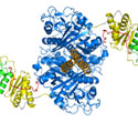
Researchers have obtained the highest-resolution image of a didomain structure
in a modular polyketide synthase (PKS), revealing new structural features. PKS
enzymes catalyze the synthesis of polyketides, which include a number of
antibiotics, anticancer agents, antiparasitics, and immunosuppressants. The
researchers solved the x-ray crystal structure of a didomain of
6-deoxyerythronolide B synthase (DEBS), a model PKS using data measured at SSRL
Structural Molecular Biology Beam Line 11-1. They imaged a 194-kDA fragment of
module 5 of the enzyme with multiwavelength anomalous dispersion (MAD). The
fragment contained full-length ketosythase (KS) and acyl transferase (AT)
domains, and the linkers that are part of the polyketide chain elongation
process. With 40,908 atoms (582 kDa) per asymmetric unit, this structure
represents the largest unique crystal structure to be solved using MAD. The
2.7-Å resolution image showed that the active site residues of the KS and AT
domains, Cys199 and Ser642, respectively, were more than 80 Å apart. The
distance is too large to be traversed by the long arm of a statically
positioned acyl carrier protein, needed to ferry growing polyketides along the
synthase backbone. The unexpected feature suggests substantial domain
reorganization may be needed for the synthase module to function. The didomain
structure also revealed a novel protein fold for the KS-to-AT linker.
Principal investigator Chaitan Khosla of Stanford University was recently
elected a fellow to the American Association for the Advancement of Science for
his contributions to the field of metabolic chemistry and engineering,
particularly to the biosynthesis of polyketide antibiotics.
To learn more about this research see the full scientific highlight at:
http://www-ssrl.slac.stanford.edu/research/highlights_archive/PKS.html
Tang, Y., Kim, C.-Y., Mathews, I. I., Cane, D. E., Khosla, C. (2006)
The 2.7-Å Crystal Structure of a 194-kDa Homodimeric Fragment of the
6-Deoxyerythronolide B Synthase. PNAS, 103, 11124-11129.