Endovascular stents made from superelastic Nitinol are a major component in the
fight against heart disease. But in order for stents to be used safely for
prolonged periods in human arteries, it is important to accurately characterize
stress/strain distributions in such stents, which govern how they deform and
fracture. SSRL scientists working at Lawrence Berkeley National Laboratory's
Advanced Light Source Beam Line 7.3.3 have taken the first direct in
situ x-ray
micro-diffraction measurements of the local strain field of a stent-like
Nitinol component subjected to realistic stresses.
Because of unique mechanical characteristics and excellent biocompatibility,
Nitinol is used as self-expanding endovascular stents to scaffold diseased
peripheral arteries. Such stents were initially designed to provide sufficient
scaffolding force to hold open vessels, yet provide enough elasticity to
withstand pulsing strains from the cardiac cycle. Many studies indicate that
these stents perform this primary function quite well. More recent in-depth
studies, however, indicate that when stents are used in peripheral arteries in
more active patients they sometime break.
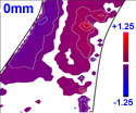
The SSRL team used a 1x1 micrometer white x-ray beam to investigate deformation
of a stent-like component under moderate to high deformation conditions.
Micro-diffraction measurements indicate that state-of-the-art commercial
finite-element models used to predict local strain fields are sufficient up to
3% deformation. However, there are significant discrepancies between measured
and calculated strains at larger displacements such as seen by superficial
femoral arteries (SFAs) as the leg is bent from an extended position. The
results show that a much better understanding of how superelastic Nitinol
accommodates high deformation is needed.
A. Mehta, X.-Y. Gong, V. Imbeni, A. R. Pelton and R. O. Ritchie, "Understanding
the Deformation and Fracture of Nitinol Endovascular Stents Using In
Situ Synchrotron X-ray Microdiffraction", Adv. Mater. 19,
1183 (2007)
To learn more about this research see the full scientific highlight at:
http://www-ssrl.slac.stanford.edu/research/highlights_archive/nitinol_stents.html