SSRL Science Highlight - October 2008

Structures and Activities Shed Light into
Cancer and Aging Phenotypes of Helicase XPD Mutations

figure 1
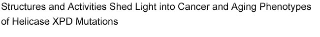
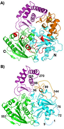
Figure 1. Ribbon diagram of crystal structures of SaXPD (A) and apo SaXPD (B). SaXPD consists of four domains: two helicase domains (cyan and green), a 4FeS domain (orange) containing a 4Fe-4S cluster (ball-sticks), and an Arch domain (purple). Seven conserved helicase motifs are highlighted in red. Disordered regions in the apoSaXPD structure are indicated by dashed lines.
Helicases are a family of enzymes universally present in all living organisms. They are involved in many DNA metabolic pathways including DNA replication, repair, recombination, transcription, translation, and RNA splicing. Consistent with their essential role in living cells, mutations in DNA helicases are associated with a variety of human genetic disorders including cancer and premature aging. The basic function of helicases is to unwind double-stranded DNA through the coupling of NTP hydrolysis with translocation along one of the two DNA strands. Most helicases contain two motor domains with RecA-like structures. XPD is a DNA helicase, first identified in xeroderma pigmentosum group D patients, and unwinds dsDNA with a 5'-3' polarity. Being part of the transcription factor TFIIH complex, XPD plays a key role in both transcription and nucleotide excision repair, a major DNA repair pathway for repairing a broad spectrum of bulky DNA damage caused by UV radiation (such as sunlight) and chemicals. Mutations in XPD are associated with three different diseases: xeroderma pigmentosum (XP), Cockayne syndrome (CS), and trichothiodystrophy (TTD). While XP patients show 1000-fold high risk of skin cancer, patients with CS or TTD have no sign of cancer predisposition but show a segmental premature aging (progeria) phenotype. A long-standing problem that scientists have tried to figure out is why mutations of the same disease are distributed cross the whole XPD polypeptide while mutations for different diseases reside closely within just a few amino acid residues. The recent X-ray crystal structures of XPD homologs determined by three independent groups, one of which led by John Tainer collected data at SSRL, have shed light into this mystery.

Using data collected at SSRL, Fan and coworkers led by John Tainer determined the crystal structure of an XPD homolog from Sulfolobus acidocaldarius (SaXPD). SaXPD is a smaller helicase than human XPD, but contains all the conserved helicase motifs and amino acid residues corresponding to 22 out of 26 disease mutations in human XPD. The crystal structure (Fig1A) reveals four domains including two motor domains (helicase domain I and II), a unique FeS domain containing a 4Fe-4S cluster with sensitivity to oxygen, and a novel Arch domain. The apo SaXPD (Fig1B) crystal structure (data collected at ALS), in which the FeS cluster was removed by soaking the crystals with oxidizing agent, indicates that the 4Fe-4S cluster is essential for the proper folding of the FeS domain and possibly plays a role in coordination of domain movements during ATP hydrolysis and DNA translocation by XPD. When mapped into the SaXPD crystal structure, the amino acid residues corresponding to 22 human diseases mutations can be distinguished into three groups matching the three different diseases (Fig2). Amino acid residues of XP mutations reside either near the ATP-binding
figure 1
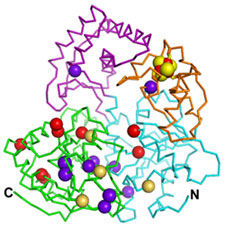
Figure 2. Distribution of amino acid residues corresponding to human XPD disease mutations. XP mutations are represented by red spheres, XP/CS mutations by greenish yellow spheres, and TTD mutations by dark purple spheres.
site at the groove formed between the two helicase motor domains or along the likely DNA interacting channel at the edge of helicase domain II. Therefore, XP mutations show reduced helicase activity. In addition to the location near active sites, amino acid residues of XP/CS mutations often show substitution of small side chains by bulky groups, and these mutations further reduce the flexibility in conformational changes required for functional enzyme activity. Different from the above two classes of disease mutations, amino acid residues of TTD mutations are distributed in all four domains. The TTD mutations likely change the protein framework of XPD, therefore possibly affect the stability of the whole enzyme and its interactions with other protein partners in the related biological processes. However, depending on its location near the active sites or not, a particular TTD mutation may or may not have direct impact on the helicase or ATP hydrolysis activity of XPD. These results provide for the first time a mechanistic explanation for these three different disease mutations, and are supported by biochemical analysis on 15 mutants carried out in the same report published in Cell magazine this May.


Primary Citation
L. Fan, J.O. Fuss, Q.J. Cheng, A.S. Arvai, M. Hammel, V.A. Roberts, P.K. Cooper and J.A. Tainer, XPD helicase structures and activities: insights into the cancer and aging phenotypes from XPD mutations, Cell 133 (2008), pp. 789-800

References

H. Liu, J. Rudolf, K.A. Johnson, S.A. McMahon, M. Oke, L. Carter, A.M. McRobbie, S.E. Brown, J.H. Naismith and M.F. White, Structure of the DNA repair helicase XPD, Cell 133 (2008), pp. 801-812.

S.C. Wolski, J. Kuper, P. Hanzelmann, J.J. Truglio, D.L. Croteau, B. Van Houten and C. Kisker, Crystal structure of the FeS cluster-containing nucleotide excision repair helicase XPD, PLoS Biol. 6 (2008), p. e149.

J. Rudolf, V. Makrantoni, W.J. Ingledew, M.J. Stark and M.F. White, The DNA repair helicases XPD and FancJ have essential iron-sulfur domains, Mol. Cell 23 (2006), pp. 801-808.

| PDF version | | Lay Summary | | Highlights Archive |


SSRL is supported by the Department of Energy, Office of Basic Energy Sciences. The SSRL Structural Molecular Biology Program is supported by the Department of Energy, Office of Biological and Environmental Research, and by the National Institutes of Health, National Center for Research Resources, Biomedical Technology Program, and the National Institute of General Medical Sciences.


Last Updated: 22 October 2008
Content Owner: L. Fan and J.A. Tainer, The Scripps Research Institute
Page Editor: L. Dunn