
Scientists studying osteoporosis and other skeletal diseases are interested in
the 3D structure of bone and its responses to conditions such as
weightlessness, radiation (of particular interest to astronauts) and vitamin D
deficiency. The current gold standard, micro-computed tomography (micro-CT),
provides 3D images of trabeculae, the small interior struts of bone tissue, and
electron microscopy can provide nanometer resolution of thin tissue slices.
Hard X-ray transmission microscopy has provided the first 3D view of bone
structure within individual trabeculae on the nanoscale.
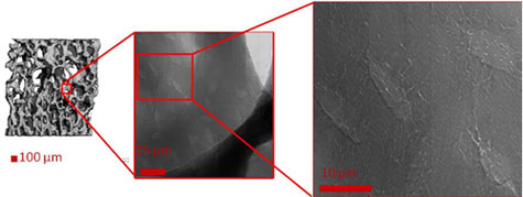
Figure 1
Micro-CT (left) shows trabecular structure inside of bone. Transmission X-ray
microscopy (TXM; center and right) can reveal localized details of osteocyte
lacunae and their processes.
Bone material responds to mechanical stresses such as experienced during
load-bearing, or its opposite weightlessness, to form new bone or break down
existing bone by complex signals. These signals are believed to be sent and
received from bone cells (osteocytes) housed within mineralized cavities called
lacunae, via cell processes that pass nutrients and other signals within
mineralized channels (canaliculi) extending from the lacunae (Duncan & Turner
1995, Khosla et al. 2008). Therefore, obtaining the 3D structure of lacunae
and canaliculi at the nanoscale will help our understanding of healthy bone
tissue and the changes that occur with aging and disease.
A team that included scientists from NASA Ames Research Center, Cornell
University, Xradia, Inc. and the Stanford Synchrotron Radiation Lightsource
(SSRL) has conducted imaging experiments of bone using the transmission x-ray
microscope (TXM) on SSRL beam line 6-2. They imaged trabeculae from mice that
were hindlimb unloaded, a method developed by NASA to simulate weightlessness,
reducing the functional loads experienced by the hindlimbs. 2D images show
detailed structures of lacunae and the associated canaliculi (Figure 1), and
tomography provided a 3D view of these structures (Figure 2, and supplemental
movie). Tomography was obtained by rotating the bony trabecula and taking
images at successive angles from -90 to 90 degrees. These images were
reconstructed mathematically to provide a 3D image, and 2D slices from the 3D
data (Fig. 2 b and c) provide detail of the lacuna and canaliculi extending
from the cell cavity.
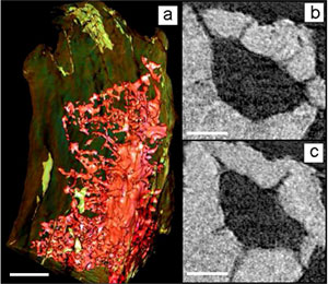
Fig. 2.
Projection (a) and slices (b and c) from tomography of single bone trabecula
imaged from -90 to 90 degrees shows lacuna and canaliculi extending from it.
The ability to image networks of osteocyte lacunae and canaliculi and to
measure local bone tissue density changes at high resolution will have
significant impact on our understanding of skeletal adaptation and disease.
This approach is already providing a novel understanding of the nanostructure
and properties of complex mineralized biological specimens and holds the
promise of being greatly informative about the nanostructure of materials and
the nanoscale complexity of life.
Primary Citation
Andrews, J.C., Almeida, E., van der Meulen, M.C.H., Alwood, J.S., Lee, C., Liu,
Y., Chen, J., Meirer, F., Feser, M., Gelb, J., Rudati, J., Tkachuk, A., Yun,
W., Pianetta, P. 2010. Nanoscale X-ray Microscopic Imaging of Mammalian
Mineralized Tissue. Microscopy and Microanalysis 16(3): 327-336.
References
Trabecular bone density was also measured quantitatively in this study, by
first calibrating TXM absorption by apatite, which is the main component of
mineralized phase in bone that absorbs X-rays. From tomographic slices of the
bone, tissue absorption was compared to that of pure apatite, to quantify
density differences within trabeculae on the nanoscale. The resolution of this
information is substantially better than can be obtained from an averaged
microscale bone density as determined by micro-CT. This nano-CT TXM method can
determine localized density changes, for example to compare newly formed vs.
older tissue after treatments to simulate and ameliorate bone disease.
SSRL is supported by the Department of Energy, Office of Basic Energy Sciences. The SSRL Structural Molecular Biology Program is supported by the Department of Energy, Office of Biological and Environmental Research, and by the National Institutes of Health, National Center for Research Resources, Biomedical Technology Program, and the National Institute of General Medical Sciences.