
3D Imaging of Bone Structure Key to Understanding Bone Health
summary written by Raven Hanna
The 3D structure of bone is critical for maintaining strength. Skeletal diseases such as osteoporosis and environmental conditions such as weightlessness, radiation, and vitamin D deficiency can affect bone structure. Understanding the 3D structure of bone is critical to understanding how these conditions affect bone's form and function.
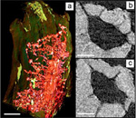
A team that included scientists from NASA Ames Research Center, Cornell University, Xradia Inc., and SSRL has developed a technique that can be used to image bone structure in 3D at high (30 nanometer) resolution. They used the transmission X ray microscope (TXM) on SSRL Beam Line 6-2 to visualize bone from mice that had undergone a process to simulate weightlessness. Using mathematically reconstructed tomography (nano-CT) images, they created a 3D image of the bone's nanostructures. The scientists were also able to measure bone density on a fine scale.
These techniques can be used to discern the difference between normal and
diseased or distressed bone, and responses to treatment. They can also
characterize nanoscale changes in bone structures due to weightlessness, and
identify differences in subsequent bone regrowth upon weight bearing. This work
is of particular interest to NASA because they are interested in the long-term
effects of weightlessness on bone density and nanostructure. This work was
published in the June issue of Microscopy and Microanalysis.
To learn more about this research see the full Scientific Highlight
Andrews, J.C., Almeida, E., van der Meulen, M.C.H., Alwood, J.S., Lee, C., Liu, Y., Chen, J., Meirer, F., Feser, M., Gelb, J., Rudati, J., Tkachuk, A., Yun, W., Pianetta, P. 2010. Nanoscale X-ray Microscopic Imaging of Mammalian Mineralized Tissue. Microscopy and Microanalysis 16(3): 327-336.
