
Visualizing Brain Metals in Health and
Disease
summary written by Raven Hanna
Metals such as iron, copper, and zinc are critical for brain function, where they serve various roles, such as enzyme cofactors or neurotransmitters. Because the metal atoms can be reactive and cause cell damage, their locations and concentrations are tightly controlled. This control can be lost in some neurodegenerative diseases. Diseases like Alzheimer's dementia and Parkinson's disease are either caused by or lead to increased metal in areas of the brain, while others, like Menkes disease, are characterized by decreased concentrations of metals. Spinocerebellar ataxia (SCA) refers to a group of degenerative disorders that cause uncoordinated movement. Some cases of SCA are linked to abnormal metal concentrations in the brain, but the specific patterns associated with various forms of the disorder are unknown.
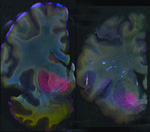
A research group led by Helen Nichol from the University of Saskatchewan used SSRL Beam Line 10-2 to compare the concentrations of iron, copper, and zinc metals in various regions of normal and SCA-affected brain tissues. The recently upgraded beam line allows measurement of the concentration of multiple metals in the same whole tissue sample using a rapid scanning X-ray fluorescence imaging technique.
In the March 24 issue of the journal Cerebellum, the researchers report areas of both higher or lower metal concentrations in the SCA brain compared to the control. Part of the basal ganglia had more iron. Part of the brainstem lacked copper. Part of the cerebellum lacked iron. Some of these results were consistent with findings from other experiments, while other observations highlight areas warranting further study. The rapid scanning XRF technique this team pioneered will be useful in future research of SCAs and other neurodegenerative diseases.
To learn more about this research see the full Scientific Highlight
Popescu, B., F., Gh, Robinson, C.A., Chapman, L.D., Nichol, H. (2009) Synchrotron X-ray Fluorescence Reveals Abnormal Metal Distributions in the Brain and Spinal Cord in Spinocerebellar Ataxia: A Case Report. Cerebellum, Mar 24 (Epub ahead of print) DOI 10.1007/s12311-009-0102-z.
