
The cells that comprise many tissues are polarized, meaning that they have
distinct 'sides' with different membrane identities. For example, in the cells
that line the intestine, the membrane that faces the space in the gut has
special proteins responsible for uptake of nutrients, whereas the sides that
contact neighboring cells have different proteins on their surfaces. Cell
polarity is fundamental to many aspects of cell and developmental biology and
it is implicated in differentiation, proliferation and morphogenesis in both
unicellular and multi-cellular organisms. Loss of cell polarity can lead to
uncontrolled tissue growth and cancers. To generate and maintain this polarized
structure, specific proteins and lipids must be delivered to particular
locations on the cell membrane. This process involves active transport of
membrane-enclosed vesicles containing specific cargo to a target site, where
the vesicle and target membranes then fuse to deliver the cargo. Soluble
N-ethylmaleimide-sensitive factor activating protein receptor (SNARE) proteins
in the vesicle membrane and target membrane form a complex that mediates
membrane fusion. The process by which SNARE-mediated membrane fusion is
coordinated with the machinery that transports the vesicle to the correct
location is poorly understood.
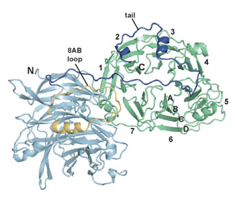 |
Figure 1:
Structure of Sro7. The N-terminal barrel is light green, the C-terminal barrel
is light blue, and the tail is shown in dark blue.
| |
In this work, Hattendorf et al. studied a yeast protein called Sro7 that is
essential for delivery of vesicles from a mother to a budding daughter cell,
which is another example of polarized cell growth. Sro7 and its relatives in
higher organisms bind to SNARE proteins and are known to be essential for cell
polarity, but their mechanism is unknown. The crystal structure of Sro7,
determined with data measured at SSRL (Beam Line 11-1) and the Advanced Light
Source, revealed a double-domain structure that is followed by a "tail" that
binds to the surface of one of the domains (Figure 1). It was shown that
removal of the tail promotes binding to a yeast SNARE protein and thereby
blocks formation of SNARE complexes. Thus, the structure led to the discovery
of an unanticipated mechanism for regulating SNARE complex assembly (Figure 2),
and provided the first mechanistic data on this essential family of proteins.
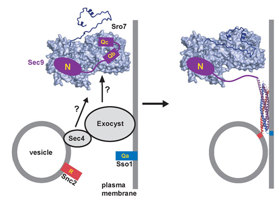 |
|
Figure 2. Model for how Sro7 may coordinate release of the SNARE Sec9
with
arrival of a secretory vesicle. Sro7 is associated with the plasma membrane at
the site of budding. It is proposed that membrane association of Sro7 is
coupled to displacement of the tail from its binding site. In this state, Sro7
binds to the Sec 9 SNARE regions (marked Qb and Qc) and prevents Sec9 from
binding to its partner SNAREs (shown in red and blue) while localizing it to
the eventual site of membrane fusion. Factors associated with the arriving
vesicle would stimulate rebinding of the tail to the body of Sro7, releasing
the Sec9 SNARE regions and allowing SNARE complex formation and membrane fusion
to proceed, thereby coordinating arrival of the vesicle with membrane
fusion. |
This work was supported by grants from the NIH and the American Cancer Society.
Primary Citation
D. A. Hattendorf, A. Andreeva, A. Gangar, P. J. Brennwald, and W. I. Weis.
(2007). Structure of the yeast polarity protein Sro7 reveals a SNARE regulatory
mechanism. Nature 446, 567-571.
References
-
Pruyne, D., Legesse-Miller, A., Gao, L., Dong, Y. & Bretscher, A.
Mechanisms of polarized growth and organelle segregation in yeast.
Annu Rev Cell Dev Biol 20, 559-91 (2004).
-
Jahn, R., Lang, T. & Südhof, T.C. Membrane fusion. Cell 112, 519-533
(2003).
|
| PDF
Version | | Lay Summary | |
Highlights Archive
|
| SSRL is supported
by the Department of Energy, Office of Basic Energy Sciences. The SSRL
Structural Molecular Biology Program is supported by the Department of Energy,
Office of Biological and Environmental Research, and by the National Institutes
of Health, National Center for Research Resources, Biomedical Technology
Program, and the National Institute of General Medical Sciences. |
|