One of the grand challenges of ultrafast science is to follow directly atomic
motion of a photo-induced reaction on the fastest time-scales and the shortest
distances—those associated with the atomic vibrations and the making and
breaking of the interatomic bonds. This is the regime that ultimately governs
chemistry and materials characteristics.
X-ray bursts produced from a free electron laser promise to be an ideal probe
to meet this challenge because of their atomic-scale structural sensitivity and
ultra-short pulse duration, which can "freeze" the atomic motion
stroboscopically [1]. However, significant technical advances
are needed before these sources can be used to make an atomic movie of the
fastest events. In particular, the optical laser pulse used to trigger the
reaction in these classes of experiments must be precisely timed with the x-ray
pulses that are used to take atomic "snap-shots".
Using the ultra-short x-ray pulses of the Sub-Picosecond Pulse Source (SPPS)
and a novel timing method, we observed the femtosecond response of a bismuth
solid following intense photoexcitation of charge carriers. Our results
provide insight into the fundamental interaction between the electronic states
and the microscopic atomic arrangements of the solid. Furthermore, we
demonstrated the ability to synchronize an optical laser to a linear
accelerator based x-ray source with femtosecond accuracy.
Bismuth is a material that shows very strong coupling between electronic and
ionic structure. It is a model system that demonstrates a rich variety of
ultrafast dynamics in the limit of high density excitations, such as extremely
large phonon amplitudes, electronic softening and phase transitions. Using
time-resolved x-ray diffraction techniques, we monitored the atomic positions
within the bismuth unit cell as a function of time in response to impulsive
photoexcitation of carriers (Figure 1). Coherent lattice oscillations were
observed similar to those previously seen in a pioneering laser plasma based
x-ray diffraction experiment [2]. However, the comparatively
large x-ray fluence of the SPPS resulted in a significant improvement in data
quality as well as enabled carrier density dependent studies.
|  |
|
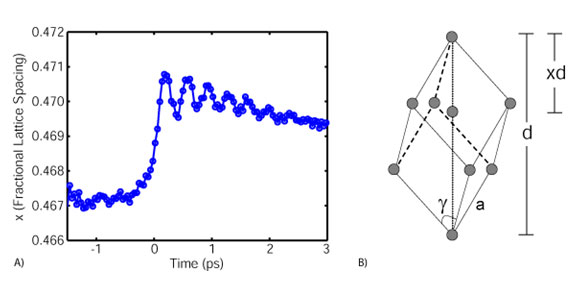
Figure 1: A) Normalized atomic coordinate as a function of time delay
for a photoexcitation fluence of 1.2 mJ/cm2. B) Crystallographic structure of
bismuth: a is the lattice constant, g is the shear angle, d is the body
diagonal, and x is the basis atom separation normalized to the body diagonal.
|
We were able to quantify the oscillation frequency and the lattice coordinate
the oscillations are occurring about from the time-resolved data. With this
information we extrapolated the curvature and minima positions of the double
well interatomic potential of bismuth as a function of photoexcited carrier
density. Our results were compared to previous density functional calculations
of the photoexcited system and are in agreement [3].
Electro-optic sampling methods were used to time the excitation laser pulse
with the x-ray probe pulse [4]. In this technique, the
electric field of the electron bunch that generated x-rays at the SPPS is used
to alter the optical properties of an electro-optic crystal (Figure 2). This
alteration is probed with a portion of the optical laser that is used to
photoexcite the bismuth sample in crossed-beam geometry. Only the portion of
the laser that is propagating within the electro-optic crystal when the
electric filed is present will be altered. In this manner, the arrival time of
the electron bunch is encoded onto spatial profile of the optical laser. The
centroid of the electro-optic feature is used to time stamp each x-ray pulse
and the data is compiled accordingly.
|  |
|
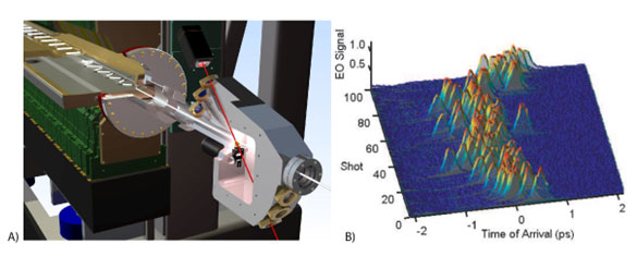
Figure 2: A) A) To time-stamp the arrival of each x-ray pulse,
researchers use an electro-optic crystal (green) placed next to the electron
beam (white) in the linear accelerator just before the beam produces x-rays. A
laser (red) probes changes in the crystal to measure the exact time the beam
passed by. The image was created by Jean Charles Castagna, SLAC. B) One
hundred consecutive electro-optic signals.
|
These measurements have furthered our understanding of bismuth dynamics far
from equilibrium. Our experiments provide the first quantitative
characterization of the curvature and quasi-equilibrium position of the
interatomic potential of a solid close to a free-carrier induced phase
transition. From this, we showed that the electronic softening of the potential
is the primary factor determining the frequency of the lattice vibrations. The
experiments also demonstrate the successful implementation of an electro-optic
timing diagnostic. This technical advancement enabled us to perform femtosecond
resolution experiments at a linear accelerator based x-ray source.
The experiments were carried out by a collaborative team from 20 different
institutions. Portions of this research were supported by the U.S. Department
of Energy, Office of Basic Energy Science through direct support for the SPPS
and the SSRL. Additional support was received by the Swedish Research Council
for Science, the Irish Research Council for Science, the Keck Foundation, the
Deutsche Forschungsgemeinschaft, the European Union RTN FLASH, the Austrian
Academy of Science, the Stanford PULSE center and the NSF FOCUS frontier
center.
Primary Citation
D. M. Fritz, D. A. Reis, B. Adams, R. A. Akre, J. Arthur, C. Blome, P. H.
Bucksbaum, A. L. Cavalieri, S. Engemann, S. Fahy, R. W. Falcone, P. H. Fuoss,
K. J. Gaffney, M. J. George, J. Hajdu, M. P. Hertlein, P. B. Hillyard, M.
Horn-von Hoegen, M. Kammler, J. Kaspar, R. Kienberger, P. Krejcik, S. H. Lee,
A. M. Lindenberg, B. McFarland, D. Meyer, T. Montagne, É. D. Murray, A. J.
Nelson, M. Nicoul, R. Pahl, J. Rudati, H. Schlarb, D. P. Siddons, K.
Sokolowski-Tinten, Th. Tschentscher, D. von der Linde and J. B. Hastings.
'Ultrafast Bond Softening in Bismuth: Mapping a Solid's Interatomic Potential
with X-rays', Science 315, 633 (2007).
References
-
R. F. Service, Science 298, 1358 (2002).
-
K. Sokolowski-Tinten, et al., Nature 422, 287 (2003).
-
É. D. Murray, et al., Phys. Rev. B 72, 060301(R) (2005).
-
A. L. Cavalieri, et al., Phys. Rev. Lett. 94, 114801 (2005).
|
Correspondence and requests for materials should be addressed to David M. Fritz
(e-mail: dmfritz@slac.stanford.edu)
| PDF
Version | | Lay Summary | |
Highlights Archive
|
| SSRL is supported
by the Department of Energy, Office of Basic Energy Sciences. The SSRL
Structural Molecular Biology Program is supported by the Department of Energy,
Office of Biological and Environmental Research, and by the National Institutes
of Health, National Center for Research Resources, Biomedical Technology
Program, and the National Institute of General Medical Sciences. |
|