|
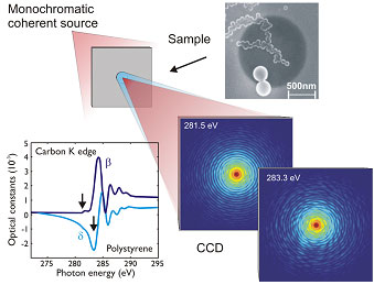 |
Figure 1: MAD imaging setup: The sample (SEM image) is illuminated with
a monochromatized and spatially coherent source (red). MAD phasing exploits the
energy-dependent interference of the resonant exit wave (red) with the
nonresonant exit wave (blue). The interference patterns recorded with a CCD
detector reveal notable changes in vicinity of the carbon K edge. The exposure
times were in the range of 700-1000 seconds with a coherent flux of
106 photons
s-1 µ-2. The corresponding optical constants for the selected energies are
indicated in the plot.
| |
|
The intricacy of structure determination due to the lost phase information in
diffraction measurements can be downsized to a manageable problem by labeling
the unknown specimen with a few heavy atoms that serve as reference scatterers.
In this way, multiple-wavelength anomalous diffraction (MAD) phasing has
revolutionized macromolecular structure determination on atomic length scales
and has become a well established technique in x-ray crystallography with
dedicated synchrotron beamlines.[1] An important prerequisite for conventional
MAD is the periodicity of the sample which requires for assembling the
biological molecules into a crystal.
In this work, the methodology of MAD is extended to non-periodic structures
using the concept of coherent x-ray imaging, cf. Figure 1. The solution of the
phase problem is demonstrated in a proof of principle experiment by a
combination of two resonantly recorded scattering or speckle patterns at the
carbon K edge. This new approach merges iterative phase retrieval
[2] and x-ray
holography approaches [3] and facilitates unique and rapid reconstructions. The
inherent resonant aspect provides sensitivity to the elemental, chemical and
magnetic state that further renders lensless MAD imaging widely applicable to a
broad range of nanostructures with in principle wavelength limited spatial
resolution.
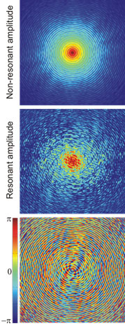 | |
Figure 2: MAD phasing results are used as reciprocal space constraints to
reconstruct the image of the specimen: (top) the nonresonant amplitudes and
(center) the resonant amplitudes of the scattered waves. Intensities are shown
on the same logarithmic scale. (bottom) The MAD phases represent the phase
relation between the two scattered waves. 800x800 pixels are shown
corresponding to a momentum transfer of 0.153 nm-1.
| |
The experiment was carried out on the soft x-ray coherent scattering Beamline
13-3 of SSRL. The speckle patterns were recorded from dispersed polysterene
spheres on a silicon nitride membrane with a gold aperture. The scattered wave
field consists out of resonant and nonresonant components and the interference
of both is recorded with an array detector in the far-field of the sample. The
strong change of the optical constants or atomic scattering factors of matter
across resonances at the threshold of core electron excitation energies allows
for defining a generalized reference wave as the energy-dependent part which
resonantly phases the nonresonant scattered wave emerging from other parts of
the sample.
The interference patterns reveal remarkable changes at nearly the same photon
energy reflecting the changes in the optical constants. The optical constants
have been determined by measuring the absorption of the sample. Given the
optical constants and applying the MAD concept the energy-dependent
interference patterns have been decomposed into resonant and non-resonant
amplitudes and their relative phases (MAD phases). The non-resonant amplitude
reproduces an Airy pattern from the circular Au aperture while the resonant
part shows a speckle pattern from the local arrangement of the polysterene
spheres.
Two images, a resonant image of the polysterene spheres and a nonresonant image
of the aperture, are simultaneously reconstructed by disentangling the MAD
|  |
|
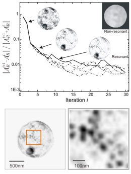
Figure 3: (top) The normalized convergence error is plotted against
the iteration. The group of convergence curves represents different
reconstructions, based on starting conditions with changes in the optical
constants by 20%. The respective reconstructions are similar within 4%
indicating the robustness of the RPR against uncertainties in the determination
of optical constants. The insets show the progressing resonant structure
determination and the nonresonant circular aperture (its simultaneous
progression is not shown). (bottom) The final resonant reconstruction of the
polysterene spheres at 25nm resolution and a magnified portion with adjusted
linear contrast.
| |
phases into resonant and nonresonant components. This is accomplished by
applying the reference-guided phase retrieval (RPR) method which has been
developed in this work. The MAD phases contain all information about the
orientation of the sample removing arbitrariness in the reconstruction process
shown in figure 3. After two iterations, the sample structure becomes apparent,
consolidates over a couple of iterations, and refines in less than 30
iterations. The resolution of MAD imaging is ultimately given by the
observation of interference terms in the speckle patterns at high momentum
transfer. In the presented case the resolution of 25nm is limited by
statistics.
Lensless MAD imaging can be viewed as an in-line x-ray holography technique
which eliminates the need for fabricating small reference structures as in
x-ray Fourier Transform Holography. In combination with the RPR algorithm the
method is capable of providing unique and rapid reconstructions of a variety of
inhomogeneous systems, representing regions with different atomic constituents,
chemical composition such as organic functional groups, and magnetic behavior
such as changes in moments and their orientations.
Primary Citation
A. Scherz, D. Zhu, R. Rick, W. F. Schlotter, S. Roy, J. Lüning, J. Stöhr,
"Nanoscale Imaging with Resonant Coherent X Rays: Extension of
Multiple-Wavelength Anomalous Diffraction to Nonperiodic Structures",
Phys. Rev. Lett. 101, 076101 (2008).
References
[1] W. A. Hendrickson, Science 254, 51 (1991).
[2] J. R. Fienup, J. Opt. Soc. Am. A 4, 118 (1987).
[3] S. Eisebitt et al., Nature (London) 432, 885 (2004).
|
|
| PDF version | | Lay
Summary | |
Highlights Archive
|
SSRL is supported
by the Department of Energy, Office of Basic Energy Sciences. The SSRL
Structural Molecular Biology Program is supported by the Department of Energy,
Office of Biological and Environmental Research, and by the National Institutes
of Health, National Center for Research Resources, Biomedical Technology
Program, and the National Institute of General Medical Sciences. |
