

Improving the quality of a high magnification image on an optical microscope is
simply a matter of cranking up the intensity of the illumination lamp. The
same is true for x-ray microscopes, but complications arise when there just
aren't enough x-rays or even worse when the sample is susceptible to damage
caused by the intense x-ray beam. To address these challenges we have
demonstrated a novel technique for improving the quality of a microscopic image
without increasing the x-ray exposure to the specimen. This affords new
opportunities to explore materials prone to soft x-ray damage, like polymer or
biological samples. Our technique uses coherent x-ray scattering to
simultaneously acquire multiple images of a specimen, which can easily be
combined later to enhance the image quality. Applying our technique in the
weak illumination limit we imaged a nanoscale test object by detecting only
2500 photons.
This holographic technique can even improve image quality under stroboscopic
illumination by an ultrafast pulsed x-ray source. Such x-ray pulses can probe
sub-picosecond dynamic processes at nanometer length scales, and will be
generated by x-ray Free Electron Lasers like the Linac Coherent Light Source
(LCLS) at SLAC.
Bright bursts of x-rays from the LCLS will not arrive until 2009. Hence, we
carried out our proof-of-principle experiment at SSRL beamline 5-2, a newly
commissioned undulator beamline optimized for soft x-ray coherent scattering.
One feature of coherent scattering is the ability to reconstruct a sample's
microscopic image from the scattering pattern alone, without using any lenses.
This can be achieved with a simple form of holography, known as Fourier
Transform Holography (FTH). (Eisebitt, et. al., Nature, 432, pp 885-887,
2004) In FTH, coherent light scattered by a sample interferes with light
scattered from a known reference aperture to form a hologram. An image of the
sample can be recovered analytically by Fourier transformation of the hologram
at a spatial resolution comparable to the size of the reference aperture.
In our example, the holographic references are defined by five nanoscale holes
that penetrate a soft x-ray opaque film as shown in Fig. 1 (a). The test
sample is a block letter F, which is little over 1 mm tall. The coherent diffraction pattern shown in Fig. 1 (b)
was recorded as a hologram with soft x-ray (l=1.58
nm) illumination of
the holographic mask. Ten reconstructed images of the sample appear in Fig. 1
(d) calculated by Fourier transformation of the hologram, but only five out of
the ten images are unique. The key element to this experiment is the
implementation of multiple holographic references. Each reference
simultaneously provides a copy of the image in the reconstruction without
increasing the exposure to the sample. These images are clear representations
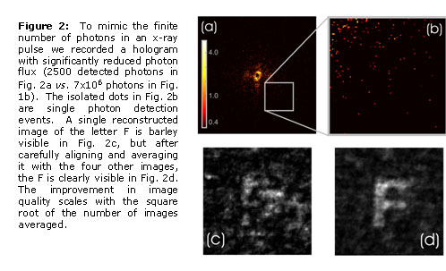
of the sample because the hologram contains ~7x10⁶ detected photons. In the
weak illumination limit, photon noise degrades the clarity as explained in Fig.
2.
Coherence, intensity and pulse structure are the hallmarks of the next
generation of x-ray light sources. With a hunger for coherence and
compatibility with pulsed illumination, lensless imaging is well suited for
these sources.
Primary Citation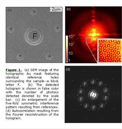
W. F. Schlotter, R. Rick, K. Chen, A. Scherz, J. Stöhr, J Lüning, S. Eisebitt,
Ch. Günther, W. Eberhardt, O. Hellwig, I. McNulty, "Multiple reference Fourier
transform holography with soft x-rays", Applied Physics Letters
89 (16): Art. No. 163112 OCT 16 2006
| PDF Version | | Lay Summary | | Highlights Archive |
| SSRL is supported by the Department of Energy, Office of Basic Energy Sciences. The SSRL Structural Molecular Biology Program is supported by the Department of Energy, Office of Biological and Environmental Research, and by the National Institutes of Health, National Center for Research Resources, Biomedical Technology Program, and the National Institute of General Medical Sciences. |