
by H. Ohldag, J. Lüning and J. Stöhr
|
by H. Ohldag, J. Lüning and J. Stöhr |
|
Spectroscopic Identification and Direct Imaging of
Interfacial Magnet Spins
Uncovering a New Layer
An der Grenze gerüttelt The determination of the crystallographic,
electronic or magnetic structure of interfaces has remained one of the great
challenges in all of materials science. The key reason is the difficulty to
detect and isolate the weak interface signature
from that of the dominant bulk. This is largely due to the
lack of depth specificity of most techniques, impeding the detection of a signal
from a well-defined depth, only. For lack of better capabilities scientists have
tried to circumvent this problem, often studying the early stages of interface
formation with surface science techniques or simply assuming "perfect"
interfaces between "bulk" materials. Using advanced soft x-ray
spectroscopy and microscopy techniques in conjunction with interface sensitive
electron yield detection we are now able to look at buried interfaces and
observe that, in reality, interfaces are quite different from model systems.
Modern magnetism is one area where interfaces play a crucial role. Todays
high-tech magnetic devices are based on thin film multilayers whose magnetic
properties depend on the magnetic coupling and spin transport across interfaces.
Examples are giant magnetoresistance structures, spin tunnel junctions, as well
as "spintronics" devices based on spin injection. A specific
interfacial problem which is of considerable scientific interest and
technological importance is the origin of "exchange bias", an effect
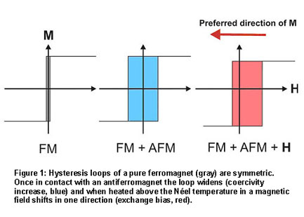
utilized in many of today's magnetic sensors and memory cells. The exchange bias effect, empirically discovered nearly 50 years ago, is used
today to create a well-defined ferromagnetic reference layer in a magnetic
device. Natural ferromagnets have a preferred magnetization "easy
axis", and an external field can align the spins into either of two equally
stable directions along this axis - the magnetization loop is symmetric as shown
in Fig. 1. When a ferromagnet (FM) is grown on an antiferromagnet (AFM) the
exchange coupling between the two systems leads to an increased coercivity of
the ferromagnet, which can be viewed as an increased "friction" to
turn the spins around. The ferromagnetic hysteresis loop is still symmetric,
indicating two equivalent easy directions. If, on the other hand, the AFM-FM
system is grown in a magnetic field or, after growth, is annealed in a magnetic
field to temperatures above the AFM Néel temperature, the hysteresis loop
becomes asymmetric and is shifted from zero, as shown in Fig. 1. This
unidirectional shift is called "exchange bias". There is now a
preferred magnetization direction for the FM along which it is most
easily aligned. The easy alignment direction can serve as a reference direction
in a device. It is
clear that exchange bias has to originate from the coupling
of the spins in the AFM to those in the FM but, because of the magnetic
neutrality of the AFM, the coupling has to involve uncompensated spins at the
AFM-FM interface. The key to the exchange bias puzzle lies in the determination
of the origin of these interfacial spins and their role in coercivity increases
and bias.
Along come two
powerful new magnetism techniques based on
soft x-rays, the x-ray magnetic circular (XMCD) and linear (XMLD) dichroism
techniques [1]. In conjunction with spectromicroscopy these x-ray techniques
have allowed a unique fresh look at the old exchange bias problem and hold the
promise to finally solving it. A series of XMCD/XMLD imaging experiments using
the Photoemission Electron Microscope (PEEM2) at the ALS previously established
the link of the AFM and FM domain structure, including the reorientation of the
AFM spins in the vicinity of the interface [2,3]. The latest, just published
[4,5], results home in on the all-important interface.
They were
obtained by two
complementary experiments, high energy resolution (150 meV) soft x-ray
absorption spectroscopy in total electron yield mode performed at Beam Line 10-1
at SSRL and high spatial resolution (50 nm) soft x-ray absorption microscopy
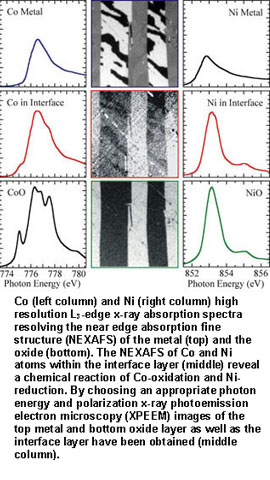
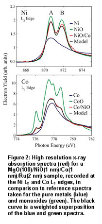
using the PEEM2 microscope at the ALS. The SSRL spectroscopy results shown in Fig. 2 demonstrate
that a thin Co layer deposited on top of bulk NiO (Co/NiO) contains Ni atoms
that are in an environment somewhere between NiO and Ni metal, and Co atoms that
are in an environment somewhere between Co metal and CoO. This is explained by an interfacial reaction in which the original NiO is reduced and the original Co
is oxidized. A new interfacial layer is formed that we shall call
NiCoOx.
References:
SSRL Highlights Archive |
 |
Last Updated: | 18 DEC 2001 |
| Content Owner: | J. Stöhr | |
| Page Editor: | Lisa Dunn |
 Computer hard drives and other advanced electronic devices depend on
layered stacks of magnetic and non-magnetic materials, but researchers
don't fully understand why such layered materials exhibit new properties
that cannot be predicted from the properties of the individual layers. In
a recent publication a team working at SSRL and the ALS describes new
methods, based on x-ray spectroscopy and x-ray microscopy, that reveal
the magnetic structures at the boundaries between these layers. Their
data show that the boundaries are not as clean as previously assumed but
a new ultrathin interface layer may be formed by a chemical reaction. The
thickness of the interfacial layer is found to change with temperature
and this change can be directly correlated with the magnetic properties
of the multilayer stack. The work provides the first magnetic images of a
buried interface and gives direct experimental evidence for the existence
and long-assumed importance of interfacial magnetic spins.
Computer hard drives and other advanced electronic devices depend on
layered stacks of magnetic and non-magnetic materials, but researchers
don't fully understand why such layered materials exhibit new properties
that cannot be predicted from the properties of the individual layers. In
a recent publication a team working at SSRL and the ALS describes new
methods, based on x-ray spectroscopy and x-ray microscopy, that reveal
the magnetic structures at the boundaries between these layers. Their
data show that the boundaries are not as clean as previously assumed but
a new ultrathin interface layer may be formed by a chemical reaction. The
thickness of the interfacial layer is found to change with temperature
and this change can be directly correlated with the magnetic properties
of the multilayer stack. The work provides the first magnetic images of a
buried interface and gives direct experimental evidence for the existence
and long-assumed importance of interfacial magnetic spins.
 It is the very difficulty associated with the determination of the
magnetic interfacial structure mentioned above, that has impeded the solution of
the exchange bias puzzle for more than forty years.
It is the very difficulty associated with the determination of the
magnetic interfacial structure mentioned above, that has impeded the solution of
the exchange bias puzzle for more than forty years.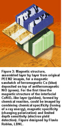 Magnetic images of the Co/NiO sandwich, obtained with the
PEEM2 microscope at the ALS, are shown in Fig. 3. The Figure is an artist's
rendition, with the original images put together layer by layer to illustrate
the magnetic structures in the NiO and Co layers and the image of the
NiCoOx.
The antiferromagnetic NiO domain image (bottom: green) was
obtained with XMLD spectromicroscopy, tuning the x-rays to the Ni L2
peaks in NiO (see Fig. 2 top). The ferromagnetic Co domain image (top: blue,
white, black) was obtained with XMCD spectromicroscopy, tuning the x-ray energy
to the Co metal L3 peak (see Fig. 2 bottom). The image of the domain
structure of the NiCoOx interface layer (middle: golden, white,
black) was obtained with XMCD spectromicroscopy, tuning the x-rays to the Ni L2
peak energy of 870.5eV (see Fig. 2 top). Since XMCD yields ferromagnetic
contrast, the observed domain structure of the interface has to arise from
uncompensated Ni spins formed by reduction of NiO. Close inspection reveals that
the domains mimic the antiferromagnetic NiO domains below and the ferromagnetic
Co domains above and therefore form the bridge between the two.
The results indicate that the increase in coercivity and the
bias shift in antiferromagnet/ferromagnet sandwiches, illustrated in Fig. 1,
originate from the interfacial spins created by a chemical reaction. Further
experiments show that in an external magnetic field most of the interfacial
spins rotate with the ferromagnet, but they provide an increased rotational
drag that leads to the widened magnetization loop. A small fraction of the
interfacial spins that has not yet been isolated, is believed
not to rotate at all in an external field because the spins are tightly
coupled to the antiferromagnetic NiO lattice underneath. These spins are
believed to give rise to the exchange bias phenomenon illustrated in Fig. 1.
Because of their small abundance, corresponding to a fraction of a monolayer,
only, the isolation of their magnetic signal and the determination of their
spatial location remain a great challenge for future experiments.
Magnetic images of the Co/NiO sandwich, obtained with the
PEEM2 microscope at the ALS, are shown in Fig. 3. The Figure is an artist's
rendition, with the original images put together layer by layer to illustrate
the magnetic structures in the NiO and Co layers and the image of the
NiCoOx.
The antiferromagnetic NiO domain image (bottom: green) was
obtained with XMLD spectromicroscopy, tuning the x-rays to the Ni L2
peaks in NiO (see Fig. 2 top). The ferromagnetic Co domain image (top: blue,
white, black) was obtained with XMCD spectromicroscopy, tuning the x-ray energy
to the Co metal L3 peak (see Fig. 2 bottom). The image of the domain
structure of the NiCoOx interface layer (middle: golden, white,
black) was obtained with XMCD spectromicroscopy, tuning the x-rays to the Ni L2
peak energy of 870.5eV (see Fig. 2 top). Since XMCD yields ferromagnetic
contrast, the observed domain structure of the interface has to arise from
uncompensated Ni spins formed by reduction of NiO. Close inspection reveals that
the domains mimic the antiferromagnetic NiO domains below and the ferromagnetic
Co domains above and therefore form the bridge between the two.
The results indicate that the increase in coercivity and the
bias shift in antiferromagnet/ferromagnet sandwiches, illustrated in Fig. 1,
originate from the interfacial spins created by a chemical reaction. Further
experiments show that in an external magnetic field most of the interfacial
spins rotate with the ferromagnet, but they provide an increased rotational
drag that leads to the widened magnetization loop. A small fraction of the
interfacial spins that has not yet been isolated, is believed
not to rotate at all in an external field because the spins are tightly
coupled to the antiferromagnetic NiO lattice underneath. These spins are
believed to give rise to the exchange bias phenomenon illustrated in Fig. 1.
Because of their small abundance, corresponding to a fraction of a monolayer,
only, the isolation of their magnetic signal and the determination of their
spatial location remain a great challenge for future experiments.