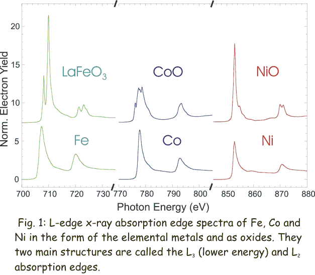
Fig. 1 (click to enlarge): L-edge x-ray absorption edge spectra of Fe, Co and Ni in the form of the elemental metals and as oxides. The two main structures are called the L3 (lower energy) and L2 absorption edges.
During the absorption process core electrons are excited into empty states above the Fermi energy and thereby probe the electronic and magnetic properties of the empty valence levels. In the following we are concerned with the spectra of the magnetic 3d transition metal elements Fe, Co and Ni. Their magnetic properties are largely determined by the 3d valence electrons. Since x-ray absorption spectra are governed by dipole selection rules the d-shell properties are best probed by L-edge absorption studies (2p to 3d transitions). The L-edge x-ray absorption spectra of the transition metals and oxides are dominated by two main peaks separated by about 15 eV as shown in Fig. 1. These two main peaks in the spectra arise from the spin orbit interaction of the 2p core shell and the total intensity of the peaks is proportional to the number of empty 3d valence states. The metal spectra mainly show two broad peaks, reflecting the width of the empty d-bands. The oxide spectra exhibit considerable fine structure, called multiplet structure. The empty oxide states are more localized than metal states and their energies are determined by crystal field and multiplet effects. Multiplet effects arise from the spin and orbital momentum coupling of different 3d valence holes (or electrons) in the electronic ground state, and from coupled states formed after x-ray absorption between the 3d valence holes and the 2p core hole.
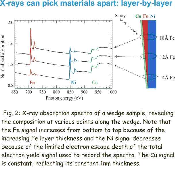
Fig. 2 (click to enlarge): X-ray absorption spectra of a wedge sample, revealing the composition at various points along the wedge. Note that the Fe signal increases from bottom to top because of the increasing Fe layer thickness. At the same time the Ni signal decreases because of the limited electron escape depth of the total electron yield signal used to record the spectra. The Cu signal is constant, reflecting its constant 1nm thickness.
There are two common approaches to detect an x-ray absorption spectra. In the first approach the intensity of an x-ray bean is measured before and after it passes through a sample using a photodiode. This method requires that the total thickness of the sample including the substrate is less than the absorption length of the x-rays which ranges between 0.1-1 micrometer. Since the x-rays travel through the entire sample and hence equally probe every atomic layer this approach provides excellent bulk sensitivity. It also excellently suited to study magnetic properties since the detected x-rays do not interact with an external magnetic field. However, very often the requirement to keep the total thickness of the sample including substrate and support is difficult to fulfill, in particular if one sets out to study single crystalline samples that need to be prepared on macroscopically thick substrates. In this case one can detect the x-ray absorption spectra by detecting the intensity of the secondary electrons that are ejected from the sample during the absorption process. This is achieved by connecting the sample to a very sensitive current amplifier and measuring the charge that flows to the sample to compensate for the charge lost during the absorption process. Figure 2 shows an example where x-ray absorption spectra were acquired in the total electron yield mode. Note, that because of the limited escape length of the detected electrons one does obtain an excellent sensitivity to the very thin Cu surface or Fe interface layer.