We all require iron, copper and zinc for normal brain function but metal metabolism becomes dysregulated in a variety of neurodegenerative diseases. Metals accumulate in Alzheimer's dementia and Parkinson's disease and are deficient in Menkes disease. Whether excess metals appear as a cause or a consequence of the disease process is not certain, but accumulation of metals have the potential to trigger cellular damage. In the healthy brain, metals are tightly regulated. Through an elegant system of copper chaperones that taxi copper from the cell surface to precise intracellular destinations, there is essentially no free copper in cells [1]. Cellular iron is not regulated by chaperones but rather by iron regulatory proteins that orchestrate and synchronize iron uptake with iron storage to reduce the availability of free iron [2]. In contrast to copper and iron, zinc is the 'wild card' of brain metal metabolism because not only does it contribute to the active site of key anti-oxidative metalloenzymes, such as the Cu-Zn superoxide dismutase, but it also exists as free zinc in some synaptic vesicles and acts as a neurotransmitter [3]. When brain tissue is damaged, such as following a stroke, free zinc can flood the injured areas resulting in cell death due to zinc excitotoxicity [4].
In an article published in the March 24 issue of Cerebellum, Popescu and co-workers have provided new insight into which brain metals are dysregulated in spinocerebellar ataxia (SCA). SCAs are a group of degenerative disorders that affect the cerebellum, the spinal cord and the connections between them causing a type of uncoordinated movement called ataxia. Friedreich's ataxia, ataxia with isolated vitamin E deficiency, X-linked sideroblastic anemia with ataxia, ataxia telangiectasia and infantile-onset spinocerebellar ataxia are examples of SCAs that have been linked to metal abnormalities. Each of these conditions has a different cause and thus, each may have a different pattern of metal dysregulation.

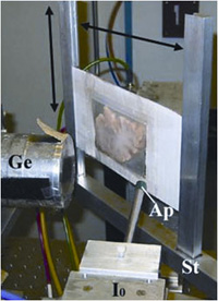
Rapid scanning X-ray fluorescence imaging [5], conducted on SSRL Beam Line 10-2, was used to compare the normal distribution of iron, copper and zinc to their distribution in a case of SCA. Formalin-fixed slices of brain and spinal cord, approximately 1 cm thick, were purchased from the Brain and Tissue Bank for Developmental Disorders at the University of Maryland, a facility funded by the National Institute of Child Health and Development. The brain and spinal cord slices were sealed without further treatment under metal-free plastic or Mylar and raster-scanned in the beam (Fig. 1). Areas of interest were subsequently excised for histological and immunohistochemical analysis.
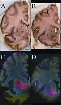
In the SCA sample, the forebrain did not show degenerative changes but the distribution of metals was unusual (Fig. 2). The basal ganglia are huge brain nuclear masses that integrate and regulate the excitatory and inhibitory circuitry adjusting the movement, amongst other functions. The globus pallidus par externa (GPe) is part of the basal ganglia involved in movement inhibition. The GPe of the SCA brain had about 80% more iron than the GPe of the normal brain. The importance of GPe iron in the development or potential treatment of SCA remains to be determined since no obvious cellular degeneration was seen in the GPe.

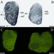
The brainstem connects the forebrain to the spinal cord. In the part of the brainstem called the medulla is an important movement coordination center named after its shape "the olive". Popescu et al. found that the nerve cell processes that pass from this region to the cerebellum are exceptionally rich in copper and this had not been previously described (Fig. 3). In contrast to the normal medulla, the 'olive' of the SCA medulla was nearly devoid of copper. It is not known why these fibers are normally rich in copper but they lack copper in SCA. However, it is interesting to note that intoxication with clioquinol, an antibiotic and potent copper chelator, causes characteristic degeneration in this brain region [6].
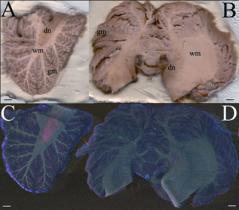
The cerebellum (Fig. 4) is quite rich in metals and contains one of the most metal-rich structures in the brain, the dentate nucleus [7]. The striking branching pattern of the cerebellar white matter called the arbor vitae (tree of life) is clearly distinguished on the basis of its metal content. In SCA the iron content of the dentate nucleus is much lower than that seen in the control brain, and the arbor vitae is not as prominent.
The high metal content of the dentate nucleus, white matter of the cerebellum and the medullary 'olive' makes these brain regions particularly susceptible to metal-catalyzed oxidative damage, neurotoxicity and neurodegeneration should metal metabolism become dysregulated. This study shows that neurodegeneration can be linked to either metal accumulation or reduction depending upon the brain region and the stage of neurodegeneration. The ability to map multiple metals in whole human brain gives us a new tool to examine metal pathologies in a variety of diseases affecting the brain and spinal cord.

Beam line 10-2 has recently been upgraded with a new set of motorized stages which allow rapid collection of x-ray fluorescence data on large samples (up to 300 mm x 600 mm) and new fluorescence x-ray digital signal processors. This new data collection system also allows rapid collection of data points, the collection of the full x-ray fluorescence spectrum at every pixel, and the ability to return to points of interest easily to further collect x-ray absorption spectra. These and other upgrades in the future will be utilized for further studies on SCAs and other neurodegenerative diseases.
- O'Halloran, T.V. and V.C. Culotta, Metallochaperones, an intracellular shuttle service for metal ions. J Biol Chem, 2000. 275(33): p. 25057-60.
- Zecca, L., et al., Iron, brain ageing and neurodegenerative disorders. Nat Rev Neurosci, 2004. 5(11): p. 863-73.
- Madsen, E. and J.D. Gitlin, Copper and iron disorders of the brain. Annu Rev Neurosci, 2007. 30: p. 317-37.
- Lee, J.M., et al., Zinc translocation accelerates infarction after mild transient focal ischemia. Neuroscience, 2002. 115(3): p. 871-8.
- Gh Popescu, B.F., et al., Mapping metals in Parkinson's and normal brain using rapid-scanning x-ray fluorescence. Phys Med Biol, 2009. 54(3): p. 651-63.
- Koga, M., A. Tsutsumi, and T. Shirabe, The pathogenesis of olivary changes in clioquinol intoxication. Neuropathology, 1997. 17: p. 290-294.
- Popescu, B.F., et al., Iron, Copper, and Zinc Distribution of the Cerebellum. Cerebellum, Jan 13. (Epub ahead of print) 2009.
Popescu, B., F., Gh, Robinson, C.A., Chapman, L.D., Nichol, H. (2009) Synchrotron X-ray Fluorescence Reveals Abnormal Metal Distributions in the Brain and Spinal Cord in Spinocerebellar Ataxia: A Case Report. Cerebellum, Mar 24 (Epub ahead of print) DOI 10.1007/s12311-009-0102-z.

