Attempting to determine and describe the atomic arrangements in an amorphous material is a daunting prospect. A considerable advance has been made in the anomalous X-ray scattering approach to determining these arrangements in materials containing two atomic species.
Up until the advent of X-ray synchrotron radiation, the X-ray radial distribution function (RDF) method was the most widely used approach for structure analysis of amorphous materials. The RDF is the probability of finding two electrons in a sample separated by a distance r, but with all the electrons of each atom positioned at the nucleus of that atom. Thus, peaks in the RDF occur at the common distances between pairs of atoms and the peak areas are determined by the number of such pairs at each distance, the coordination numbers. The RDF is readily interpreted for an amorphous material containing only one atomic species, like amorphous Se. For many samples containing more than one atomic species, however, the RDF cannot be interpreted unambiguously, as different pairs of atomic species can have almost the same interatomic distance and contribute to the same peak.
The advent of synchrotron radiation made it possible to obtain more definitive structural information via analysis of Extended X-ray Absorption Fine Structure (EXAFS). Under the best of circumstances, one can determine the average surrounding of each atomic species in the sample. That is, one can determine the average distances to its neighbors, the number of such neighbors and the atomic species of the neighbors. Unfortunately, this ideal situation is often not achieved because one cannot apply the analysis to the EXAFS at energies less than some tens of volts above the absorption edge. As a consequence, information about next near neighbors and beyond is often lost and the information about nearest neighbors is unreliable if there is a broad spread in interatomic distances - a characteristic of many amorphous metallic and semiconducting alloys (in contrast to the situation in strongly covalently bonded organic systems).
The Differential Anomalous X-ray Scattering (DAXS) technique, developed at SSRL over two decades, has provided a valuable complement to EXAFS. This approach, like EXAFS, provides information about the coordination of a specific atomic species, using scattering data obtained at two photon energies just below the absorption edge of the atomic species of interest. In contrast to EXAFS, scattering data are readily obtained at the low scattering vectors corresponding to the missing region from the EXAFS data. Thus, it provides considerably more information about next near neighbors and beyond, as well as systems with a broad spread in near neighbor distances. Generally, however, it cannot obtain information at large scattering vectors that are accessible to EXAFS. As a consequence, it often cannot resolve two different, almost equal interatomic distances. For a two component system αβ, if the α-α, α-β and β-bdistances are close, the method cannot determine whether the neighbors of an α are α or β atoms - or some combination of αs and βs.
This latter shortcoming of anomalous X-ray scattering (AXS) has now been overcome. It has been known for a very long time that one can, in principle, distinguish between α-α, β-β and α-β neighbors with AXS by taking measurements at two photon energies just below the a edge and just below the b edge, as well as at one photon energy far from both edges[1]. From these data, one obtains α-β partial pair distribution functions (PPDFs) which give the number of β atoms surrounding an a atom at a distance r. The problem has been the extreme sensitivity of these PPDFs to experimental errors and, particularly, uncertainties in the inelastic scattering which must be subtracted from the measured intensities.
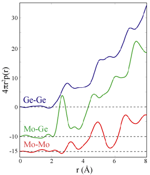
What Ishii et al. have done to overcome this limitation is to use a diffracted beam analyzer to eliminate the inelastic scattering experimentally[2]. This analyzer consists of a sagitally-focusing graphite monochromator which disperses the energies emanating from the sample onto a position-sensitive detector [3,4]. Thus at each scattering angle, an energy spectrum is measured with elastic, fluorescent and Compton peaks separately resolved. These data have resulted in a dramatic improvement in the quality of the derived PPDFs. The sample studied, amorphous MoGe3, is of interest because earlier work had suggested that MoGe3 is the metal-rich endpoint for phase separation in the sputtered Mo-Ge amorphous alloy system[5]. This is unexpected, as the first crystalline compound found in the equilibrium Mo-Ge system is MoGe2. Thus one important question for this study was whether a new compound is formed in the sputter-deposited alloys. The primary results of this study are shown in Figure 1, which shows the Ge-Ge, Ge-Mo, and Mo-Mo PPDFs. Analysis of the Mo-Ge PPDF implies an eight-fold coordination of Mo by Ge - a conclusion unobtainable by EXAFS. Further, there is no evidence of phase-separation. Although not exploited in this paper, there is clearly additional information in the higher shells of the PPDFs due to the lower starting scattering vector.
These results highlight the great promise of AXS for the study of amorphous materials, especially binary alloys.
- A. G. Munro, Phys Rev. B 25, 5037 (1982).
- H. Ishii, S. Brennan and A. Bienenstock, J. Non-Cryst. Sol. 299-302, 243 (2002).
- G. E. Ice, C. J. Sparks, Nucl. Instrum. Meth A 291, 110 (1990).
- A. K. Freund, A. Munkholm, S. Brennan, SPIE Proc 2856, 68 (1996).
- M. J. Regan, A. Bienenstock, Phys. Rev. B 51 12170 (1995).


