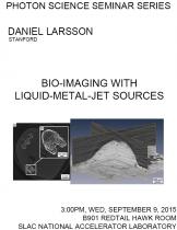
Speaker: Daniel Larsson, Stanford
Program Description
In this talk, I present results from 2D and 3D imaging experiments using liquid-metal-jet sources [1,2]. These high-brightness laboratory sources typically operate at 40 W e-beam power and ~7 µm x-ray spot size, enabling high-resolution absorption imaging and in-line phase contrast for improved soft-tissue visibility.
I will give an overview of recent bio-imaging applications investigated by my group at KTH in Stockholm, such as mouse tomography and tumor demarcation [3], phase-contrast micro‑angiography in mouse ears [4] and zebrafish muscle imaging [5].
I also show first imaging results with a new high-power source [6] (Excillum AB, Sweden) which has recently been commissioned in our lab. At present, it constitutes an improvement in brightness by a factor of six compared to previous liquid-metal-jet sources.References
[1] Hemberg O. et al., Appl. Phys. Lett. 83 (7), 1483-1485 (2003).
[2] Larsson D. H. et al., Rev. Sci. Instrum. 82, 123701 (2011).
[3] Larsson D. H. et al., Med. Phys. 40, 021909-021915 (2013).
[4] Lundström U. et al., Phys. Med. Biol. 59 2801–2811 (2014).
[5] Vagberg W. & Larsson D. H. et al., in press.
[6] Larsson D. H. et al., manuscript in preparation.


